 Movie
Movie Controller


+ Open data

- Basic information
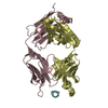
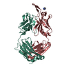
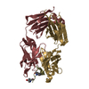
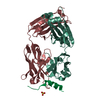
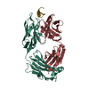
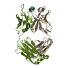
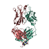
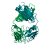
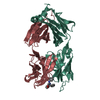
| Entry | Database: PDB / ID: 1nlb | ||||||
|---|---|---|---|---|---|---|---|
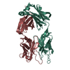
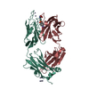
| Title | crystal structure of anti-HCV monoclonal antibody 19D9D6 | ||||||
 Components Components |
| ||||||
 Keywords Keywords | IMMUNE SYSTEM / murine monoclonal antibody | ||||||
| Function / homology | Immunoglobulins / Immunoglobulin-like / Sandwich / Mainly Beta Function and homology information Function and homology information | ||||||
| Biological species |  | ||||||
| Method |  X-RAY DIFFRACTION / SYNCHROTRON / MOLECULAR REPLACEMENT / Resolution: 1.6 Å X-RAY DIFFRACTION / SYNCHROTRON / MOLECULAR REPLACEMENT / Resolution: 1.6 Å | ||||||
 Authors Authors | Menez, R. / Bossus, M. / Muller, B. / Sibai, G. / Dalbon, P. / Ducancel, F. / Jolivet-Reynaud, C. / Stura, E. | ||||||
 Citation Citation | Journal: J.Immunol. / Year: 2003 Title: Crystal structure of a hydrophobic immunodominant antigenic site on hepatitis C virus core protein complexed to monoclonal antibody 19D9D6. Authors: Menez, R. / Bossus, M. / Muller, B.H. / Sibai, G. / Dalbon, P. / Ducancel, F. / Jolivet-Reynaud, C. / Stura, E.A. | ||||||
| History |
|
- Structure visualization
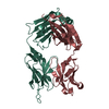
| Structure viewer | Molecule: MolmilJmol/JSmol |
|---|
- Downloads & links
-Download
| PDBx/mmCIF format | 1nlb.cif.gz | 113.6 KB | Display | PDBx/mmCIF format |
|---|---|---|---|---|
| PDB format | pdb1nlb.ent.gz | 86.4 KB | Display | PDB format |
| PDBx/mmJSON format | 1nlb.json.gz | Tree view | PDBx/mmJSON format | |
| Others |  Other downloads Other downloads |
-Validation report
| Arichive directory | https://data.pdbj.org/pub/pdb/validation_reports/nl/1nlbftp://data.pdbj.org/pub/pdb/validation_reports/nl/1nlb | HTTPS FTP |
|---|
-Related structure data
| Related structure data |  1n64SC S: Starting model for refinement C: citing same article ( |
|---|---|
| Similar structure data |


-Links
 PDBj
PDBj

- Assembly
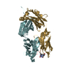
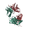
| Deposited unit | 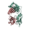
| ||||||||
|---|---|---|---|---|---|---|---|---|---|
| 1 |
| ||||||||
| Unit cell |
| ||||||||
| Components on special symmetry positions |
|
-Components
| #1: Antibody | Mass: 24334.051 Da / Num. of mol.: 1 / Source method: isolated from a natural source / Source: (natural) |
|---|---|
| #2: Antibody | Mass: 23563.430 Da / Num. of mol.: 1 / Source method: isolated from a natural source / Source: (natural) |
| #3: Water | ChemComp-HOH /  Mass: 18.015 Da / Num. of mol.: 715 / Source method: isolated from a natural source / Formula: H2O Mass: 18.015 Da / Num. of mol.: 715 / Source method: isolated from a natural source / Formula: H2O |
| Has protein modification | Y |
-Experimental details
-Experiment
| Experiment | Method: X-RAY DIFFRACTION / Number of used crystals: 1 |
|---|
- Sample preparation
| Crystal | Density Matthews: 2.4 Å3/Da / Density % sol: 48.75 % |
|---|---|
| Crystal grow | Temperature: 290 K / Method: vapor diffusion, sitting drop / pH: 9 Details: 12.5% MPEG 5000, 250mM NaCl, 100mM tris/HCl, pH 9, VAPOR DIFFUSION, SITTING DROP, temperature 290K |
| Crystal grow | *PLUS Method: unknown / Details: Stura, E.A., (2001) J. Crystal Growth, 232, 545. |
-Data collection
| Diffraction | Mean temperature: 100 K |
|---|---|
| Diffraction source | Source: SYNCHROTRON / Site: ESRF  / Beamline: ID14-2 / Wavelength: 0.933 / Beamline: ID14-2 / Wavelength: 0.933 |
| Detector | Type: ADSC QUANTUM 4 / Detector: CCD / Date: Nov 11, 2001 / Details: mirrors |
| Radiation | Monochromator: diamond(111), Ge(220) / Protocol: SINGLE WAVELENGTH / Monochromatic (M) / Laue (L): M / Scattering type: x-ray |
| Radiation wavelength | Wavelength: 0.933 Å / Relative weight: 1 |
| Reflection | Resolution: 1.6→20 Å / Num. all: 47973 / Num. obs: 42445 / % possible obs: 79.5 % / Observed criterion σ(I): 0 |
| Reflection shell | Resolution: 1.6→1.66 Å / % possible all: 42 |
| Reflection | *PLUS Lowest resolution: 20 Å / Num. measured all: 47973 / Rmerge(I) obs: 0.08 |
| Reflection shell | *PLUS % possible obs: 42 % / Rmerge(I) obs: 0.22 / Mean I/σ(I) obs: 1.5 |
- Processing
| Software |
| ||||||||||||||||||||
|---|---|---|---|---|---|---|---|---|---|---|---|---|---|---|---|---|---|---|---|---|---|
| Refinement | Method to determine structure: MOLECULAR REPLACEMENT Starting model: PDB 1N64 Resolution: 1.6→20 Å / σ(F): 2 / σ(I): 0 / Stereochemistry target values: Engh & Huber
| ||||||||||||||||||||
| Refinement step | Cycle: LAST / Resolution: 1.6→20 Å
| ||||||||||||||||||||
| Refine LS restraints |
| ||||||||||||||||||||
| LS refinement shell | Resolution: 1.6→1.66 Å /
| ||||||||||||||||||||
| Refinement | *PLUS Lowest resolution: 20 Å | ||||||||||||||||||||
| Solvent computation | *PLUS | ||||||||||||||||||||
| Displacement parameters | *PLUS | ||||||||||||||||||||
| LS refinement shell | *PLUS Rfactor Rfree: 0.235 / Rfactor Rwork: 0.2 |
