 Movie
Movie Controller

[English] 日本語
 Yorodumi
- PDB-1nc3: Crystal structure of E. coli MTA/AdoHcy nucleosidase complexed wi... -
+ Open data

- Basic information
| Entry | Database: PDB / ID: 1nc3 | ||||||
|---|---|---|---|---|---|---|---|
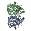
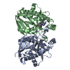
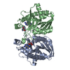
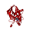
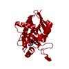
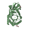
| Title | Crystal structure of E. coli MTA/AdoHcy nucleosidase complexed with formycin A (FMA) | ||||||
 Components Components | MTA/SAH nucleosidase | ||||||
 Keywords Keywords | HYDROLASE / mixed alpha/beta dimer | ||||||
| Function / homology |  Function and homology information Function and homology informationpurine deoxyribonucleoside catabolic process / toxic metabolite repair / adenosylhomocysteine nucleosidase / adenosylhomocysteine nucleosidase activity / methylthioadenosine nucleosidase activity / : / : / protein homodimerization activity / identical protein binding / cytosol Similarity search - Function | ||||||
| Biological species |  | ||||||
| Method |  X-RAY DIFFRACTION / MOLECULAR REPLACEMENT / Resolution: 2.2 Å X-RAY DIFFRACTION / MOLECULAR REPLACEMENT / Resolution: 2.2 Å | ||||||
 Authors Authors | Lee, J.E. / Cornell, K.A. / Riscoe, M.K. / Howell, P.L. | ||||||
 Citation Citation | Journal: J.Biol.Chem. / Year: 2003 Title: Structure of Escherichia coli 5'-methylthioadenosine/ S-adenosylhomocysteine nucleosidase inhibitor complexes provide insight into the conformational changes required for substrate binding and catalysis. Authors: Lee, J.E. / Cornell, K.A. / Riscoe, M.K. / Howell, P.L. | ||||||
| History |
|
- Structure visualization
| Structure viewer | Molecule: MolmilJmol/JSmol |
|---|
- Downloads & links
-Download
| PDBx/mmCIF format | 1nc3.cif.gz | 101.1 KB | Display | PDBx/mmCIF format |
|---|---|---|---|---|
| PDB format | pdb1nc3.ent.gz | 77.6 KB | Display | PDB format |
| PDBx/mmJSON format | 1nc3.json.gz | Tree view | PDBx/mmJSON format | |
| Others |  Other downloads Other downloads |
-Validation report
| Arichive directory | https://data.pdbj.org/pub/pdb/validation_reports/nc/1nc3ftp://data.pdbj.org/pub/pdb/validation_reports/nc/1nc3 | HTTPS FTP |
|---|
-Related structure data
| Related structure data |  1nc1C  1jysS C: citing same article ( S: Starting model for refinement |
|---|---|
| Similar structure data |
-Links
 PDBj
PDBj- Assembly
| Deposited unit | 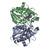
| ||||||||
|---|---|---|---|---|---|---|---|---|---|
| 1 |
| ||||||||
| Unit cell |
|
-Components
| #1: Protein | Mass: 25488.123 Da / Num. of mol.: 2 Source method: isolated from a genetically manipulated source Source: (gene. exp.) References: UniProt: P24247, UniProt: P0AF12*PLUS, adenosylhomocysteine nucleosidase #2: Chemical |   Mass: 267.241 Da / Num. of mol.: 2 / Source method: obtained synthetically / Formula: C10H13N5O4 Mass: 267.241 Da / Num. of mol.: 2 / Source method: obtained synthetically / Formula: C10H13N5O4#3: Water | ChemComp-HOH / |  Mass: 18.015 Da / Num. of mol.: 200 / Source method: isolated from a natural source / Formula: H2O Mass: 18.015 Da / Num. of mol.: 200 / Source method: isolated from a natural source / Formula: H2ONonpolymer details | Waters 1-98 are associated with chain A and waters 99-200 are associated with chain B. | |
|---|
-Experimental details
-Experiment
| Experiment | Method: X-RAY DIFFRACTION / Number of used crystals: 1 |
|---|
- Sample preparation
| Crystal | Density Matthews: 2.23 Å3/Da / Density % sol: 44.88 % | |||||||||||||||||||||||||||||||||||||||||||||||||
|---|---|---|---|---|---|---|---|---|---|---|---|---|---|---|---|---|---|---|---|---|---|---|---|---|---|---|---|---|---|---|---|---|---|---|---|---|---|---|---|---|---|---|---|---|---|---|---|---|---|---|
| Crystal grow | Temperature: 293 K / Method: vapor diffusion, hanging drop / pH: 4.7 Details: PEG 200, sodium acetate, sodium chloride, cobalt chloride, pH 4.7, VAPOR DIFFUSION, HANGING DROP, temperature 293K | |||||||||||||||||||||||||||||||||||||||||||||||||
| Crystal grow | *PLUS | |||||||||||||||||||||||||||||||||||||||||||||||||
| Components of the solutions | *PLUS
|
-Data collection
| Diffraction | Mean temperature: 100 K |
|---|---|
| Diffraction source | Source: ROTATING ANODE / Type: RIGAKU RU300 / Wavelength: 1.5418 Å |
| Detector | Type: RIGAKU RAXIS IV++ / Detector: IMAGE PLATE / Date: Jan 12, 2001 / Details: Confocal multilayer |
| Radiation | Monochromator: Confocal multilayer / Protocol: SINGLE WAVELENGTH / Monochromatic (M) / Laue (L): M / Scattering type: x-ray |
| Radiation wavelength | Wavelength: 1.5418 Å / Relative weight: 1 |
| Reflection | Resolution: 2.2→47.48 Å / Num. obs: 23876 / % possible obs: 99.9 % / Observed criterion σ(F): 0 / Observed criterion σ(I): 0 / Redundancy: 5.6 % / Rmerge(I) obs: 0.072 |
| Reflection shell | Resolution: 2.2→2.34 Å / Redundancy: 5.6 % / Rmerge(I) obs: 0.22 / Num. unique all: 3920 / % possible all: 99.8 |
| Reflection | *PLUS Highest resolution: 2.2 Å / Lowest resolution: 50 Å / Num. obs: 29777 / % possible obs: 99.8 % / Redundancy: 5.7 % / Num. measured all: 170060 / Rmerge(I) obs: 0.077 |
| Reflection shell | *PLUS Lowest resolution: 2.3 Å / % possible obs: 99.5 % / Rmerge(I) obs: 0.261 / Mean I/σ(I) obs: 2.7 |
- Processing
| Software |
| |||||||||||||||||||||||||
|---|---|---|---|---|---|---|---|---|---|---|---|---|---|---|---|---|---|---|---|---|---|---|---|---|---|---|
| Refinement | Method to determine structure: MOLECULAR REPLACEMENT Starting model: PDB code: 1JYS Resolution: 2.2→47.48 Å / Isotropic thermal model: Overall / Cross valid method: THROUGHOUT / σ(F): 0 / σ(I): 0 / Stereochemistry target values: Engh & Huber
| |||||||||||||||||||||||||
| Displacement parameters | Biso mean: 29.8 Å2
| |||||||||||||||||||||||||
| Refine analyze |
| |||||||||||||||||||||||||
| Refinement step | Cycle: LAST / Resolution: 2.2→47.48 Å
| |||||||||||||||||||||||||
| Refine LS restraints |
| |||||||||||||||||||||||||
| LS refinement shell | Resolution: 2.2→2.34 Å / Rfactor Rfree error: 0.022
| |||||||||||||||||||||||||
| Refinement | *PLUS Highest resolution: 2.2 Å / Lowest resolution: 50 Å / % reflection Rfree: 5 % | |||||||||||||||||||||||||
| Solvent computation | *PLUS | |||||||||||||||||||||||||
| Displacement parameters | *PLUS | |||||||||||||||||||||||||
| Refine LS restraints | *PLUS
|
