 Movie
Movie Controller

[English] 日本語
 Yorodumi
- PDB-1nbu: 7,8-Dihydroneopterin Aldolase Complexed with Product From Mycobac... -
+ Open data

- Basic information
| Entry | Database: PDB / ID: 1nbu | ||||||
|---|---|---|---|---|---|---|---|
| Title | 7,8-Dihydroneopterin Aldolase Complexed with Product From Mycobacterium Tuberculosis | ||||||
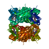
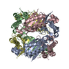
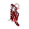
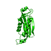
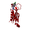
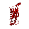
 Components Components | (Probable dihydroneopterin aldolase) x 3 | ||||||
 Keywords Keywords | LYASE / anti-parallel / beta-sheet / two alpha helices / Structural Genomics / PSI / Protein Structure Initiative / TB Structural Genomics Consortium / TBSGC | ||||||
| Function / homology |  Function and homology information Function and homology information7,8-dihydroneopterin oxygenase / 7,8-dihydroneopterin epimerase / dihydroneopterin aldolase / dihydroneopterin aldolase activity / encapsulin nanocompartment / folic acid biosynthetic process / isomerase activity / tetrahydrofolate biosynthetic process / oxidoreductase activity / cytoplasm Similarity search - Function | ||||||
| Biological species |   Mycobacterium tuberculosis (bacteria) Mycobacterium tuberculosis (bacteria) | ||||||
| Method |  X-RAY DIFFRACTION / AB INITIO / Resolution: 1.6 Å X-RAY DIFFRACTION / AB INITIO / Resolution: 1.6 Å | ||||||
 Authors Authors | Goulding, C.W. / Apostol, M.I. / Sawaya, M.R. / Phillips, M. / Parseghian, A. / Eisenberg, D. / TB Structural Genomics Consortium (TBSGC) | ||||||
 Citation Citation | Journal: J.Mol.Biol. / Year: 2005 Title: Regulation by oligomerization in a mycobacterial folate biosynthetic enzyme. Authors: Goulding, C.W. / Apostol, M.I. / Sawaya, M.R. / Phillips, M. / Parseghian, A. / Eisenberg, D. | ||||||
| History |
|
- Structure visualization
| Structure viewer | Molecule: MolmilJmol/JSmol |
|---|
- Downloads & links
-Download
| PDBx/mmCIF format | 1nbu.cif.gz | 204 KB | Display | PDBx/mmCIF format |
|---|---|---|---|---|
| PDB format | pdb1nbu.ent.gz | 163.1 KB | Display | PDB format |
| PDBx/mmJSON format | 1nbu.json.gz | Tree view | PDBx/mmJSON format | |
| Others |  Other downloads Other downloads |
-Validation report
| Arichive directory | https://data.pdbj.org/pub/pdb/validation_reports/nb/1nbuftp://data.pdbj.org/pub/pdb/validation_reports/nb/1nbu | HTTPS FTP |
|---|
-Related structure data
| Related structure data |  1z9wC  1dhnS S: Starting model for refinement C: citing same article ( |
|---|---|
| Similar structure data | |
| Other databases |

-Links
 PDBj
PDBj
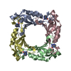
- Assembly
| Deposited unit | 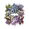
| ||||||||
|---|---|---|---|---|---|---|---|---|---|
| 1 |
| ||||||||
| Unit cell |
|
-Components
| #1: Protein | Mass: 13251.920 Da / Num. of mol.: 5 Source method: isolated from a genetically manipulated source Source: (gene. exp.) Mycobacterium tuberculosis (bacteria) / Gene: FOLB OR RV3607C OR MT3712.1 OR MTCY07H7B.15 / Plasmid: pET22b+ / Species (production host): Escherichia coli / Production host: References: UniProt: P0A580, UniProt: P9WNC5*PLUS, dihydroneopterin aldolase #2: Protein | Mass: 13207.910 Da / Num. of mol.: 2 / Mutation: D19A Source method: isolated from a genetically manipulated source Source: (gene. exp.) Mycobacterium tuberculosis (bacteria) / Gene: FOLB OR RV3607C OR MT3712.1 OR MTCY07H7B.15 / Production host: References: UniProt: P0A580, UniProt: P9WNC5*PLUS, dihydroneopterin aldolase #3: Protein | | Mass: 13115.813 Da / Num. of mol.: 1 / Mutation: Y18A, D19A Source method: isolated from a genetically manipulated source Source: (gene. exp.) Mycobacterium tuberculosis (bacteria) / Gene: FOLB OR RV3607C OR MT3712.1 OR MTCY07H7B.15 / Production host: References: UniProt: P0A580, UniProt: P9WNC5*PLUS, dihydroneopterin aldolase #4: Chemical | ChemComp-PH2 /   Mass: 195.179 Da / Num. of mol.: 8 / Source method: obtained synthetically / Formula: C7H9N5O2 Mass: 195.179 Da / Num. of mol.: 8 / Source method: obtained synthetically / Formula: C7H9N5O2#5: Water | ChemComp-HOH / |  Mass: 18.015 Da / Num. of mol.: 470 / Source method: isolated from a natural source / Formula: H2O Mass: 18.015 Da / Num. of mol.: 470 / Source method: isolated from a natural source / Formula: H2O |
|---|
-Experimental details
-Experiment
| Experiment | Method: X-RAY DIFFRACTION / Number of used crystals: 1 |
|---|
- Sample preparation
| Crystal | Density Matthews: 1.73 Å3/Da / Density % sol: 29 % | |||||||||||||||||||||||||||||||||||||||||||||||||
|---|---|---|---|---|---|---|---|---|---|---|---|---|---|---|---|---|---|---|---|---|---|---|---|---|---|---|---|---|---|---|---|---|---|---|---|---|---|---|---|---|---|---|---|---|---|---|---|---|---|---|
| Crystal grow | Temperature: 300 K / Method: vapor diffusion, hanging drop / pH: 4.6 Details: PEG 4000, sodium acetate, pH 4.6, VAPOR DIFFUSION, HANGING DROP, temperature 300K | |||||||||||||||||||||||||||||||||||||||||||||||||
| Crystal grow | *PLUS pH: 7.4 / Method: vapor diffusion, hanging drop | |||||||||||||||||||||||||||||||||||||||||||||||||
| Components of the solutions | *PLUS
|
-Data collection
| Diffraction | Mean temperature: 200 K |
|---|---|
| Diffraction source | Source: ROTATING ANODE / Type: RIGAKU / Wavelength: 1.5418 Å |
| Detector | Type: RIGAKU RAXIS IV / Detector: IMAGE PLATE / Date: Jan 17, 2002 / Details: mirrors |
| Radiation | Monochromator: yale mirrors / Protocol: SINGLE WAVELENGTH / Monochromatic (M) / Laue (L): M / Scattering type: x-ray |
| Radiation wavelength | Wavelength: 1.5418 Å / Relative weight: 1 |
| Reflection | Resolution: 1.6→90 Å / Num. all: 118185 / Num. obs: 118185 / % possible obs: 99.2 % / Observed criterion σ(F): 1 / Observed criterion σ(I): 1 / Rmerge(I) obs: 0.068 |
| Reflection shell | Resolution: 1.6→1.66 Å / Rmerge(I) obs: 0.464 / % possible all: 93.9 |
| Reflection | *PLUS Lowest resolution: 100 Å / Num. obs: 118216 / Rmerge(I) obs: 0.069 |
| Reflection shell | *PLUS % possible obs: 93.9 % / Num. unique obs: 111905 / Rmerge(I) obs: 0.5 / Mean I/σ(I) obs: 1.9 |
- Processing
| Software |
| |||||||||||||||||||||||||||||||||
|---|---|---|---|---|---|---|---|---|---|---|---|---|---|---|---|---|---|---|---|---|---|---|---|---|---|---|---|---|---|---|---|---|---|---|
| Refinement | Method to determine structure: AB INITIO Starting model: PDB ENTRY 1DHN Resolution: 1.6→10 Å / Num. parameters: 71829 / Num. restraintsaints: 90056 / Cross valid method: FREE R / σ(F): 0 / Stereochemistry target values: ENGH AND HUBER Details: ANISOTROPIC REFINEMENT REDUCED FREE R (NO CUTOFF), Author states ARG23 of chain A, ARG23 of chain B, ARG15 of chain C, ARG15 of chain E, ARG23 of chain E and ARG23 of chain F all have two distinct conformations.
| |||||||||||||||||||||||||||||||||
| Refine analyze | Num. disordered residues: 0 / Occupancy sum hydrogen: 0 / Occupancy sum non hydrogen: 7947.25 | |||||||||||||||||||||||||||||||||
| Refinement step | Cycle: LAST / Resolution: 1.6→10 Å
| |||||||||||||||||||||||||||||||||
| Refine LS restraints |
| |||||||||||||||||||||||||||||||||
| Software | *PLUS Name: SHELXL / Version: 97 / Classification: refinement | |||||||||||||||||||||||||||||||||
| Refinement | *PLUS Highest resolution: 1.6 Å / Lowest resolution: 20 Å / Rfactor Rfree: 0.258 / Rfactor Rwork: 0.165 | |||||||||||||||||||||||||||||||||
| Solvent computation | *PLUS | |||||||||||||||||||||||||||||||||
| Displacement parameters | *PLUS | |||||||||||||||||||||||||||||||||
| Refine LS restraints | *PLUS
|
