 Movie
Movie Controller

[English] 日本語
 Yorodumi
- PDB-1dhn: 1.65 ANGSTROM RESOLUTION STRUCTURE OF 7,8-DIHYDRONEOPTERIN ALDOLA... -
+ Open data

- Basic information
| Entry | Database: PDB / ID: 1dhn | ||||||
|---|---|---|---|---|---|---|---|
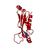
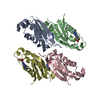
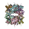
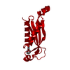
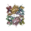
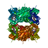
| Title | 1.65 ANGSTROM RESOLUTION STRUCTURE OF 7,8-DIHYDRONEOPTERIN ALDOLASE FROM STAPHYLOCOCCUS AUREUS | ||||||
 Components Components | 7,8-DIHYDRONEOPTERIN ALDOLASE | ||||||
 Keywords Keywords | PTERIN BINDING / FOLATE BIOSYNTHESIS / ANTIBIOTIC TARGET / BETA-BARREL | ||||||
| Function / homology |  Function and homology information Function and homology information7,8-dihydroneopterin epimerase / dihydroneopterin aldolase / dihydroneopterin aldolase activity / folic acid biosynthetic process / isomerase activity / tetrahydrofolate biosynthetic process / cytoplasm Similarity search - Function | ||||||
| Biological species |   Staphylococcus aureus (bacteria) Staphylococcus aureus (bacteria) | ||||||
| Method |  X-RAY DIFFRACTION / MIR / Resolution: 1.65 Å X-RAY DIFFRACTION / MIR / Resolution: 1.65 Å | ||||||
 Authors Authors | Hennig, M. / D'Arcy, A. / Hampele, I.C. / Page, M.G.P. / Oefner, C.H. / Dale, G. | ||||||
 Citation Citation | Journal: Nat.Struct.Biol. / Year: 1998 Title: Crystal structure and reaction mechanism of 7,8-dihydroneopterin aldolase from Staphylococcus aureus. Authors: Hennig, M. / D'Arcy, A. / Hampele, I.C. / Page, M.G. / Oefner, C. / Dale, G.E. | ||||||
| History |
|
- Structure visualization
| Structure viewer | Molecule: MolmilJmol/JSmol |
|---|
- Downloads & links
-Download
| PDBx/mmCIF format | 1dhn.cif.gz | 37.4 KB | Display | PDBx/mmCIF format |
|---|---|---|---|---|
| PDB format | pdb1dhn.ent.gz | 26.4 KB | Display | PDB format |
| PDBx/mmJSON format | 1dhn.json.gz | Tree view | PDBx/mmJSON format | |
| Others |  Other downloads Other downloads |
-Validation report
| Arichive directory | https://data.pdbj.org/pub/pdb/validation_reports/dh/1dhnftp://data.pdbj.org/pub/pdb/validation_reports/dh/1dhn | HTTPS FTP |
|---|
-Related structure data


-Links
 PDBj
PDBj
- Assembly
| Deposited unit | 
| ||||||||
|---|---|---|---|---|---|---|---|---|---|
| 1 | x 8
| ||||||||
| Unit cell |
|
-Components
| #1: Protein | Mass: 13769.635 Da / Num. of mol.: 1 Source method: isolated from a genetically manipulated source Details: OCTAMER IS CRYSTALLOGRAPHIC / Source: (gene. exp.) Staphylococcus aureus (bacteria) / Gene: DHNA / Plasmid: PSADHNA / Production host: |
|---|---|
| #2: Water | ChemComp-HOH /  Mass: 18.015 Da / Num. of mol.: 122 / Source method: isolated from a natural source / Formula: H2O Mass: 18.015 Da / Num. of mol.: 122 / Source method: isolated from a natural source / Formula: H2O |
-Experimental details
-Experiment
| Experiment | Method: X-RAY DIFFRACTION / Number of used crystals: 1 |
|---|
- Sample preparation
| Crystal | Density Matthews: 2.1 Å3/Da / Density % sol: 41.92 % Description: STRUCTURE SOLVED BY SE-MET AND K2PTCL4 DERIVATIVES | |||||||||||||||
|---|---|---|---|---|---|---|---|---|---|---|---|---|---|---|---|---|
| Crystal grow | pH: 6.5 / Details: pH 6.5 | |||||||||||||||
| Crystal grow | *PLUS Method: vapor diffusion, hanging drop | |||||||||||||||
| Components of the solutions | *PLUS
|
-Data collection
| Diffraction | Mean temperature: 120 K |
|---|---|
| Diffraction source | Source: ROTATING ANODE / Type: ENRAF-NONIUS FR591 / Wavelength: 1.5418 |
| Detector | Type: MARRESEARCH / Detector: IMAGE PLATE / Date: Sep 4, 1996 / Details: SUPPER MIRROR |
| Radiation | Monochromatic (M) / Laue (L): M / Scattering type: x-ray |
| Radiation wavelength | Wavelength: 1.5418 Å / Relative weight: 1 |
| Reflection | Resolution: 1.65→30 Å / Num. obs: 14310 / % possible obs: 98.4 % / Observed criterion σ(I): 0 / Redundancy: 3.9 % / Biso Wilson estimate: 22.5 Å2 / Rmerge(I) obs: 0.047 / Net I/σ(I): 12.4 |
| Reflection | *PLUS Lowest resolution: 30 Å / Num. measured all: 56369 |
- Processing
| Software |
| ||||||||||||||||||||||||||||||||||||||||||||||||||||||||||||
|---|---|---|---|---|---|---|---|---|---|---|---|---|---|---|---|---|---|---|---|---|---|---|---|---|---|---|---|---|---|---|---|---|---|---|---|---|---|---|---|---|---|---|---|---|---|---|---|---|---|---|---|---|---|---|---|---|---|---|---|---|---|
| Refinement | Method to determine structure: MIR / Resolution: 1.65→30 Å / Data cutoff low absF: 0 / Cross valid method: RFREE / σ(F): 0
| ||||||||||||||||||||||||||||||||||||||||||||||||||||||||||||
| Displacement parameters | Biso mean: 27.4 Å2 | ||||||||||||||||||||||||||||||||||||||||||||||||||||||||||||
| Refinement step | Cycle: LAST / Resolution: 1.65→30 Å
| ||||||||||||||||||||||||||||||||||||||||||||||||||||||||||||
| Refine LS restraints |
| ||||||||||||||||||||||||||||||||||||||||||||||||||||||||||||
| Xplor file |
| ||||||||||||||||||||||||||||||||||||||||||||||||||||||||||||
| Software | *PLUS Name: X-PLOR / Classification: refinement | ||||||||||||||||||||||||||||||||||||||||||||||||||||||||||||
| Refinement | *PLUS % reflection Rfree: 5 % | ||||||||||||||||||||||||||||||||||||||||||||||||||||||||||||
| Solvent computation | *PLUS | ||||||||||||||||||||||||||||||||||||||||||||||||||||||||||||
| Displacement parameters | *PLUS |
