 Movie
Movie Controller


+ Open data

- Basic information
| Entry | Database: PDB / ID: 1n7d | |||||||||
|---|---|---|---|---|---|---|---|---|---|---|
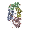
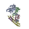
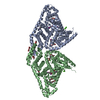
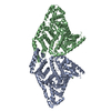
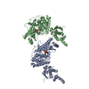
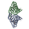
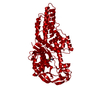
| Title | Extracellular domain of the LDL receptor | |||||||||
 Components Components | Low-density lipoprotein receptor | |||||||||
 Keywords Keywords | LIPID TRANSPORT / LDL-Receptor / familial hypercholesterolemia / LDL / cholesterol metabolism | |||||||||
| Function / homology |  Function and homology information Function and homology informationreceptor-mediated endocytosis involved in cholesterol transport / regulation of phosphatidylcholine catabolic process / plasma lipoprotein particle clearance / negative regulation of astrocyte activation / positive regulation of lysosomal protein catabolic process / negative regulation of microglial cell activation / very-low-density lipoprotein particle receptor activity / PCSK9-LDLR complex / low-density lipoprotein particle clearance / cholesterol import ...receptor-mediated endocytosis involved in cholesterol transport / regulation of phosphatidylcholine catabolic process / plasma lipoprotein particle clearance / negative regulation of astrocyte activation / positive regulation of lysosomal protein catabolic process / negative regulation of microglial cell activation / very-low-density lipoprotein particle receptor activity / PCSK9-LDLR complex / low-density lipoprotein particle clearance / cholesterol import / positive regulation of triglyceride biosynthetic process / negative regulation of receptor recycling / intestinal cholesterol absorption / clathrin heavy chain binding / Chylomicron clearance / low-density lipoprotein particle receptor activity / amyloid-beta clearance by cellular catabolic process / cholesterol transport / low-density lipoprotein particle binding / LDL clearance / high-density lipoprotein particle clearance / phospholipid transport / response to caloric restriction / regulation of protein metabolic process / low-density lipoprotein particle / artery morphogenesis / negative regulation of amyloid fibril formation / negative regulation of protein metabolic process / cellular response to fatty acid / endolysosome membrane / negative regulation of low-density lipoprotein particle clearance / regulation of cholesterol metabolic process / sorting endosome / lipoprotein particle binding / amyloid-beta clearance / cellular response to low-density lipoprotein particle stimulus / cholesterol metabolic process / long-term memory / phagocytosis / Retinoid metabolism and transport / cholesterol homeostasis / retinoid metabolic process / somatodendritic compartment / clathrin-coated pit / receptor-mediated endocytosis / lipid metabolic process / clathrin-coated endocytic vesicle membrane / endocytosis / positive regulation of inflammatory response / apical part of cell / late endosome / Cargo recognition for clathrin-mediated endocytosis / amyloid-beta binding / Clathrin-mediated endocytosis / virus receptor activity / protease binding / molecular adaptor activity / basolateral plasma membrane / early endosome / signaling receptor complex / lysosome / endosome membrane / negative regulation of gene expression / external side of plasma membrane / positive regulation of gene expression / calcium ion binding / Golgi apparatus / cell surface / membrane / identical protein binding / plasma membrane Similarity search - Function | |||||||||
| Biological species |  Homo sapiens (human) Homo sapiens (human) | |||||||||
| Method |  X-RAY DIFFRACTION / SYNCHROTRON / MAD / Resolution: 3.7 Å X-RAY DIFFRACTION / SYNCHROTRON / MAD / Resolution: 3.7 Å | |||||||||
 Authors Authors | Rudenko, G. / Henry, L. / Henderson, K. / Ichtchenko, K. / Brown, M.S. / Goldstein, J.L. / Deisenhofer, J. | |||||||||
 Citation Citation | Journal: Science / Year: 2002 Title: Structure of the LDL receptor extracellular domain at endosomal pH Authors: Rudenko, G. / Henry, L. / Henderson, K. / Ichtchenko, K. / Brown, M.S. / Goldstein, J.L. / Deisenhofer, J. #1: Journal: Acta Crystallogr.,Sect.D / Year: 2003 Title: 'MAD'ly phasing the extracellular domain of the LDL receptor: a medium-sized protein, large tungsten clusters and multiple non-isomorphous crystals. Authors: Rudenko, G. / Henry, L. / Vonrhein, C. / Bricogne, G. / Deisenhofer, J. | |||||||||
| History |
| |||||||||
| Remark 300 | BIOMOLECULE: 1 THIS ENTRY CONTAINS THE CRYSTALLOGRAPHIC ASYMMETRIC UNIT WHICH CONSISTS OF 1 CHAIN(S) ...BIOMOLECULE: 1 THIS ENTRY CONTAINS THE CRYSTALLOGRAPHIC ASYMMETRIC UNIT WHICH CONSISTS OF 1 CHAIN(S). SEE REMARK 350 FOR INFORMATION ON GENERATING THE BIOLOGICAL MOLECULE(S). THE AUTHORS BELIEVE THE BIOLOGICAL UNIT IS A MONOMER. | |||||||||
| Remark 600 | HETEROGEN THE ATOMS W1 AND W2 OF THE LIGAND KEG 6003 ARE ON A TWO-FOLD AXIS. COORDINATES FOR THE ...HETEROGEN THE ATOMS W1 AND W2 OF THE LIGAND KEG 6003 ARE ON A TWO-FOLD AXIS. COORDINATES FOR THE HALF OF THE CLUSTER KEG 6003 IN THE ASYMMETRIC UNIT ARE INCLUDED IN THIS ENTRY. THE SECOND HALF OF THE CLUSTER CAN BE GENERATED USING SYMMETRY OPERATION Y,X,1-Z. |
- Structure visualization
| Structure viewer | Molecule: MolmilJmol/JSmol |
|---|
- Downloads & links
-Download
| PDBx/mmCIF format | 1n7d.cif.gz | 136.3 KB | Display | PDBx/mmCIF format |
|---|---|---|---|---|
| PDB format | pdb1n7d.ent.gz | 97.1 KB | Display | PDB format |
| PDBx/mmJSON format | 1n7d.json.gz | Tree view | PDBx/mmJSON format | |
| Others |  Other downloads Other downloads |
-Validation report
| Arichive directory | https://data.pdbj.org/pub/pdb/validation_reports/n7/1n7dftp://data.pdbj.org/pub/pdb/validation_reports/n7/1n7d | HTTPS FTP |
|---|
-Related structure data
| Related structure data |  1ajjS  1d2jS 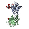 1fx5S  1i0uS  1ijqS S: Starting model for refinement |
|---|---|
| Similar structure data |

-Links
 PDBj
PDBj


- Assembly
| Deposited unit | 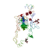
| ||||||||||||||||||
|---|---|---|---|---|---|---|---|---|---|---|---|---|---|---|---|---|---|---|---|
| 1 |
| ||||||||||||||||||
| Unit cell |
| ||||||||||||||||||
| Components on special symmetry positions |
|
-Components
| #1: Protein | Mass: 77703.617 Da / Num. of mol.: 1 / Fragment: extracellular domain, RESIDUES 1-699 / Mutation: N494Q, N636Q Source method: isolated from a genetically manipulated source Source: (gene. exp.) Homo sapiens (human) / Gene: LDLR / Production host:  Trichoplusia ni (cabbage looper) / References: UniProt: P01130 Trichoplusia ni (cabbage looper) / References: UniProt: P01130 | ||||
|---|---|---|---|---|---|
| #2: Polysaccharide | alpha-D-mannopyranose-(1-3)-[alpha-D-mannopyranose-(1-6)]beta-D-mannopyranose-(1-4)-2-acetamido-2- ...alpha-D-mannopyranose-(1-3)-[alpha-D-mannopyranose-(1-6)]beta-D-mannopyranose-(1-4)-2-acetamido-2-deoxy-beta-D-glucopyranose-(1-4)-2-acetamido-2-deoxy-beta-D-glucopyranose Source method: isolated from a genetically manipulated source | ||||
| #3: Polysaccharide | alpha-D-mannopyranose-(1-6)-beta-D-mannopyranose-(1-4)-2-acetamido-2-deoxy-beta-D-glucopyranose-(1- ...alpha-D-mannopyranose-(1-6)-beta-D-mannopyranose-(1-4)-2-acetamido-2-deoxy-beta-D-glucopyranose-(1-4)-2-acetamido-2-deoxy-beta-D-glucopyranose Source method: isolated from a genetically manipulated source | ||||
| #4: Chemical | ChemComp-CA /   Mass: 40.078 Da / Num. of mol.: 8 / Source method: obtained synthetically / Formula: Ca Mass: 40.078 Da / Num. of mol.: 8 / Source method: obtained synthetically / Formula: Ca#5: Chemical |   Mass: 2877.030 Da / Num. of mol.: 3 / Source method: obtained synthetically / Formula: O40PW12 Mass: 2877.030 Da / Num. of mol.: 3 / Source method: obtained synthetically / Formula: O40PW12Has protein modification | Y | |
-Experimental details
-Experiment
| Experiment | Method: X-RAY DIFFRACTION / Number of used crystals: 8 |
|---|
- Sample preparation
| Crystal | Density Matthews: 5.43 Å3/Da / Density % sol: 77.36 % | ||||||||||||||||||||||||||||||||||||||||||
|---|---|---|---|---|---|---|---|---|---|---|---|---|---|---|---|---|---|---|---|---|---|---|---|---|---|---|---|---|---|---|---|---|---|---|---|---|---|---|---|---|---|---|---|
| Crystal grow | Temperature: 298 K / Method: vapor diffusion, hanging drop / pH: 5.3 Details: acetate buffer pH 5.3, 1,2-hexanediol, 0.5 mM CaCl2, VAPOR DIFFUSION, HANGING DROP, temperature 298K | ||||||||||||||||||||||||||||||||||||||||||
| Crystal | *PLUS Density % sol: 74 % | ||||||||||||||||||||||||||||||||||||||||||
| Crystal grow | *PLUS Method: unknown | ||||||||||||||||||||||||||||||||||||||||||
| Components of the solutions | *PLUS
|
-Data collection
| Diffraction |
| ||||||||||||||||||||
|---|---|---|---|---|---|---|---|---|---|---|---|---|---|---|---|---|---|---|---|---|---|
| Diffraction source |
| ||||||||||||||||||||
| Detector |
| ||||||||||||||||||||
| Radiation | Protocol: MAD / Monochromatic (M) / Laue (L): M / Scattering type: x-ray | ||||||||||||||||||||
| Radiation wavelength |
| ||||||||||||||||||||
| Reflection | Resolution: 3.7→45.3 Å / Num. obs: 17303 / % possible obs: 95.6 % / Observed criterion σ(F): 0 / Observed criterion σ(I): 0 / Redundancy: 3.7 % / Biso Wilson estimate: 74.7 Å2 / Rmerge(I) obs: 0.09 / Net I/σ(I): 9.8 | ||||||||||||||||||||
| Reflection shell | Resolution: 3.7→3.83 Å / Rmerge(I) obs: 0.449 / Mean I/σ(I) obs: 2.7 / % possible all: 64.5 | ||||||||||||||||||||
| Reflection | *PLUS Lowest resolution: 33.7 Å / Num. measured all: 64801 / Rmerge(I) obs: 0.09 | ||||||||||||||||||||
| Reflection shell | *PLUS Highest resolution: 3.7 Å / % possible obs: 64.5 % |

- Processing
| Software |
| |||||||||||||||||||||||||
|---|---|---|---|---|---|---|---|---|---|---|---|---|---|---|---|---|---|---|---|---|---|---|---|---|---|---|
| Refinement | Method to determine structure: MAD Starting model: PDB ENTRIES: 1AJJ, 1D2J, 1I0U, 1IJQ, 1FX5 Resolution: 3.7→45.3 Å / Rfactor Rfree error: 0.011 / Isotropic thermal model: OVERALL / Cross valid method: THROUGHOUT / σ(F): 0 / Stereochemistry target values: Engh & Huber Details: The sugars have not been refined, just manually placed in the density. The exact identity of the sugars in these N-linked glycosylation sites is not known. At this resolution positional ...Details: The sugars have not been refined, just manually placed in the density. The exact identity of the sugars in these N-linked glycosylation sites is not known. At this resolution positional refinement is of limited use. Harmonic restraints were used on most core residues. No simulated annealing was used. The resolution of the MAD data used in the structure determination is 3.7 Ang and the B-factors were high. This means that the positions of the side chains are not well determined in the model. The authors urge users to exercize caution and restraint interpreting this model. The authors are available for questions. The clusters were included in the positional refinement (regularization of the model). The carbohydrates were not included in the positional refinement (regularization of the model) but fitted manually. The carbohydrates were modelled as the N-linked high mannose type.
| |||||||||||||||||||||||||
| Solvent computation | Solvent model: FLAT MODEL / Bsol: 10 Å2 / ksol: 0.156563 e/Å3 | |||||||||||||||||||||||||
| Displacement parameters | Biso mean: 124.7 Å2
| |||||||||||||||||||||||||
| Refine analyze |
| |||||||||||||||||||||||||
| Refinement step | Cycle: LAST / Resolution: 3.7→45.3 Å
| |||||||||||||||||||||||||
| Refine LS restraints |
| |||||||||||||||||||||||||
| LS refinement shell | Resolution: 3.7→3.87 Å / Rfactor Rfree error: 0.062 / Total num. of bins used: 8
| |||||||||||||||||||||||||
| Xplor file |
| |||||||||||||||||||||||||
| Refinement | *PLUS Highest resolution: 3.7 Å / Lowest resolution: 33.1 Å / % reflection Rfree: 7 % / Rfactor Rfree: 0.392 / Rfactor Rwork: 0.388 | |||||||||||||||||||||||||
| Solvent computation | *PLUS | |||||||||||||||||||||||||
| Displacement parameters | *PLUS | |||||||||||||||||||||||||
| Refine LS restraints | *PLUS
|
