 Movie
Movie Controller


+ Open data

- Basic information
| Entry | Database: PDB / ID: 1fx5 | |||||||||
|---|---|---|---|---|---|---|---|---|---|---|
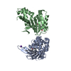
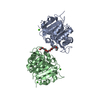
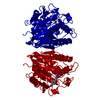
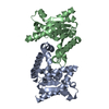
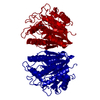
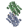
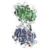
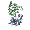
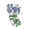
| Title | CRYSTAL STRUCTURE ANALYSIS OF ULEX EUROPAEUS LECTIN I | |||||||||
 Components Components | ANTI-H(O) LECTIN I | |||||||||
 Keywords Keywords | SUGAR BINDING PROTEIN / Legume lectin / UE-I / homo-dimer / fucose specific lectin | |||||||||
| Function / homology |  Function and homology information Function and homology information | |||||||||
| Biological species |  Ulex europaeus (furze) Ulex europaeus (furze) | |||||||||
| Method |  X-RAY DIFFRACTION / MOLECULAR REPLACEMENT / Resolution: 2.2 Å X-RAY DIFFRACTION / MOLECULAR REPLACEMENT / Resolution: 2.2 Å | |||||||||
 Authors Authors | Audette, G.F. / Vandonselaar, M. / Delbaere, L.T.J. | |||||||||
 Citation Citation | Journal: J.Mol.Biol. / Year: 2000 Title: The 2.2 A resolution structure of the O(H) blood-group-specific lectin I from Ulex europaeus. Authors: Audette, G.F. / Vandonselaar, M. / Delbaere, L.T. | |||||||||
| History |
|
- Structure visualization
| Structure viewer | Molecule: MolmilJmol/JSmol |
|---|
- Downloads & links
-Download
| PDBx/mmCIF format | 1fx5.cif.gz | 116.5 KB | Display | PDBx/mmCIF format |
|---|---|---|---|---|
| PDB format | pdb1fx5.ent.gz | 88.4 KB | Display | PDB format |
| PDBx/mmJSON format | 1fx5.json.gz | Tree view | PDBx/mmJSON format | |
| Others |  Other downloads Other downloads |
-Validation report
| Arichive directory | https://data.pdbj.org/pub/pdb/validation_reports/fx/1fx5ftp://data.pdbj.org/pub/pdb/validation_reports/fx/1fx5 | HTTPS FTP |
|---|
-Related structure data
| Related structure data | 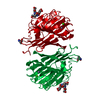 1lecS S: Starting model for refinement |
|---|---|
| Similar structure data |

-Links
 PDBj
PDBj

- Assembly
| Deposited unit | 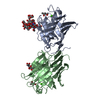
| ||||||||||
|---|---|---|---|---|---|---|---|---|---|---|---|
| 1 |
| ||||||||||
| Unit cell |
| ||||||||||
| Noncrystallographic symmetry (NCS) | NCS oper: (Code: given Matrix: (-0.9953, 0.09681, 0.00399), Vector: Details | Biological assembly is a homo-dimer. The deposited structure differs from other legume lectins with the presence of a disulfide bond between Cys 115 and Cys 150. The Ala 86 of the Ala-Asp cis-peptide fequently observed in legume lectins is a Thr in UE-I. Residues Glu 28, Lys 83, Asn 240, Thr 241 of chain A and Glu 28, Asn 39, Asn 40, Lys 83, Lys 114, and Asn 240 of chain B were refined as Ala's due to lack of electron density for these side-chains. Several residues differ from the published primary sequence on the basis of the observed electron density. Asn 116 of both chains A and B could not be accomodated in the observed electron density, and was removed; however, residue numbering from 117 to 243 was retained. The final two (2) C-terminal amino acid residues in chain A and final three (3) C-terminal amino acid residues of chain B were not observed in the electron density. | |
-Components
-Protein , 1 types, 2 molecules AB
| #1: Protein | Mass: 26629.494 Da / Num. of mol.: 2 / Source method: isolated from a natural source Details: THERE IS AN N-LINKED HEPTASACCHARIDE AT ASN 23 OF CHAIN A AND AN N-LINKED TRISACCHARIDE AT ASN 111 OF CHAINS A AND B Source: (natural) Ulex europaeus (furze) / Organ: SEEDS / References: UniProt: P22972 |
|---|
-Sugars , 2 types, 3 molecules
| #2: Polysaccharide | Xylitol-(1-2)-[alpha-D-mannopyranose-(1-3)][alpha-D-mannopyranose-(1-6)]beta-D-mannopyranose-(1-4)- ...Xylitol-(1-2)-[alpha-D-mannopyranose-(1-3)][alpha-D-mannopyranose-(1-6)]beta-D-mannopyranose-(1-4)-2-acetamido-2-deoxy-beta-D-glucopyranose-(1-4)-[alpha-L-fucopyranose-(1-3)]2-acetamido-2-deoxy-beta-D-glucopyranose Type: oligosaccharide / Mass: 1191.095 Da / Num. of mol.: 1 Source method: isolated from a genetically manipulated source |
|---|---|
| #3: Polysaccharide | Source method: isolated from a genetically manipulated source |
-Non-polymers , 4 types, 200 molecules 


| #4: Chemical |  Mass: 54.938 Da / Num. of mol.: 2 / Source method: obtained synthetically / Formula: Mn Mass: 54.938 Da / Num. of mol.: 2 / Source method: obtained synthetically / Formula: Mn#5: Chemical |  Mass: 40.078 Da / Num. of mol.: 2 / Source method: obtained synthetically / Formula: Ca Mass: 40.078 Da / Num. of mol.: 2 / Source method: obtained synthetically / Formula: Ca#6: Chemical |  Mass: 118.174 Da / Num. of mol.: 2 / Source method: obtained synthetically / Formula: C6H14O2 / Comment: precipitant*YM Mass: 118.174 Da / Num. of mol.: 2 / Source method: obtained synthetically / Formula: C6H14O2 / Comment: precipitant*YM#7: Water | ChemComp-HOH / | Mass: 18.015 Da / Num. of mol.: 194 / Source method: isolated from a natural source / Formula: H2O |
|---|
-Details
| Has protein modification | Y |
|---|
-Experimental details
-Experiment
| Experiment | Method: X-RAY DIFFRACTION / Number of used crystals: 3 |
|---|
- Sample preparation
| Crystal | Density Matthews: 3.06 Å3/Da / Density % sol: 59.75 % | ||||||||||||||||||||||||||||||||||||||||||||||||
|---|---|---|---|---|---|---|---|---|---|---|---|---|---|---|---|---|---|---|---|---|---|---|---|---|---|---|---|---|---|---|---|---|---|---|---|---|---|---|---|---|---|---|---|---|---|---|---|---|---|
| Crystal grow | Temperature: 287 K / Method: vapor diffusion, sitting drop / pH: 4 Details: R,S-2-methyl-2,4-pentanediol, sodium acetate, MnCl2, CaCl2, sodium cacodylate, pH 4.0, VAPOR DIFFUSION, SITTING DROP, temperature 287.0K | ||||||||||||||||||||||||||||||||||||||||||||||||
| Crystal grow | *PLUS | ||||||||||||||||||||||||||||||||||||||||||||||||
| Components of the solutions | *PLUS
|
-Data collection
| Diffraction |
| ||||||||||||||||
|---|---|---|---|---|---|---|---|---|---|---|---|---|---|---|---|---|---|
| Diffraction source |
| ||||||||||||||||
| Detector |
| ||||||||||||||||
| Radiation | Protocol: SINGLE WAVELENGTH / Monochromatic (M) / Laue (L): M / Scattering type: x-ray | ||||||||||||||||
| Radiation wavelength | Wavelength: 1.5418 Å / Relative weight: 1 | ||||||||||||||||
| Reflection | Resolution: 2.2→40 Å / Num. all: 29087 / Num. obs: 29087 / % possible obs: 89.2 % / Observed criterion σ(I): 2 / Redundancy: 2.1 % / Biso Wilson estimate: 22.5 Å2 / Rmerge(I) obs: 0.056 / Net I/σ(I): 9.5 | ||||||||||||||||
| Reflection shell | Resolution: 2.2→2.32 Å / Redundancy: 1.2 % / Rmerge(I) obs: 0.105 / Num. unique all: 3015 / % possible all: 63.6 | ||||||||||||||||
| Reflection | *PLUS | ||||||||||||||||
| Reflection shell | *PLUS % possible obs: 63.9 % |
- Processing
| Software |
| |||||||||||||||||||||||||
|---|---|---|---|---|---|---|---|---|---|---|---|---|---|---|---|---|---|---|---|---|---|---|---|---|---|---|
| Refinement | Method to determine structure: MOLECULAR REPLACEMENT Starting model: Griffonia simplicifolia lectin IV, 1LEC Resolution: 2.2→40 Å / Cross valid method: THROUGHOUT / σ(F): 0 / σ(I): 0 / Stereochemistry target values: Engh & Huber / Details: Simulated Annealing
| |||||||||||||||||||||||||
| Refinement step | Cycle: LAST / Resolution: 2.2→40 Å
| |||||||||||||||||||||||||
| Refine LS restraints |
| |||||||||||||||||||||||||
| Software | *PLUS Name: X-PLOR / Version: 3.1 / Classification: refinement | |||||||||||||||||||||||||
| Refinement | *PLUS Highest resolution: 2.2 Å / Lowest resolution: 40 Å / Num. reflection obs: 26189 / σ(F): 0 / % reflection Rfree: 10 % | |||||||||||||||||||||||||
| Solvent computation | *PLUS | |||||||||||||||||||||||||
| Displacement parameters | *PLUS | |||||||||||||||||||||||||
| Refine LS restraints | *PLUS
|
