 Movie
Movie Controller

[English] 日本語
 Yorodumi
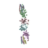
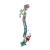
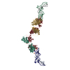
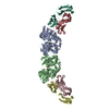
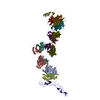
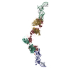
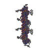
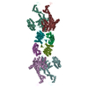
- PDB-1n73: Fibrin D-Dimer, Lamprey complexed with the PEPTIDE LIGAND: GLY-HI... -
+ Open data

- Basic information
| Entry | Database: PDB / ID: 1n73 | ||||||
|---|---|---|---|---|---|---|---|
| Title | Fibrin D-Dimer, Lamprey complexed with the PEPTIDE LIGAND: GLY-HIS-ARG-PRO-AMIDE | ||||||
 Components Components |
| ||||||
 Keywords Keywords | BLOOD CLOTTING / isopeptide cross-linked chains / protein-peptide complex | ||||||
| Function / homology |  Function and homology information Function and homology informationblood coagulation, common pathway / induction of bacterial agglutination / fibrinogen complex / platelet alpha granule / Regulation of TLR by endogenous ligand / cellular response to leptin stimulus / MyD88 deficiency (TLR2/4) / positive regulation of heterotypic cell-cell adhesion / IRAK4 deficiency (TLR2/4) / MyD88:MAL(TIRAP) cascade initiated on plasma membrane ...blood coagulation, common pathway / induction of bacterial agglutination / fibrinogen complex / platelet alpha granule / Regulation of TLR by endogenous ligand / cellular response to leptin stimulus / MyD88 deficiency (TLR2/4) / positive regulation of heterotypic cell-cell adhesion / IRAK4 deficiency (TLR2/4) / MyD88:MAL(TIRAP) cascade initiated on plasma membrane / plasminogen activation / extracellular matrix structural constituent / p130Cas linkage to MAPK signaling for integrins / positive regulation of peptide hormone secretion / GRB2:SOS provides linkage to MAPK signaling for Integrins / positive regulation of exocytosis / blood coagulation, fibrin clot formation / protein polymerization / negative regulation of endothelial cell apoptotic process / positive regulation of vasoconstriction / cellular response to interleukin-1 / Integrin cell surface interactions / : / negative regulation of extrinsic apoptotic signaling pathway via death domain receptors / fibrinolysis / Integrin signaling / positive regulation of substrate adhesion-dependent cell spreading / platelet alpha granule lumen / cell-matrix adhesion / positive regulation of protein secretion / Signaling by high-kinase activity BRAF mutants / MAP2K and MAPK activation / response to calcium ion / platelet aggregation / Signaling by RAF1 mutants / Signaling by moderate kinase activity BRAF mutants / Paradoxical activation of RAF signaling by kinase inactive BRAF / Signaling downstream of RAS mutants / Signaling by BRAF and RAF1 fusions / Platelet degranulation / extracellular vesicle / protein-folding chaperone binding / extracellular matrix / ER-Phagosome pathway / protein-containing complex assembly / blood microparticle / cell cortex / adaptive immune response / positive regulation of ERK1 and ERK2 cascade / protein-macromolecule adaptor activity / external side of plasma membrane / signaling receptor binding / innate immune response / synapse / structural molecule activity / cell surface / endoplasmic reticulum / : / extracellular exosome / extracellular region / metal ion binding / plasma membrane Similarity search - Function | ||||||
| Biological species |  | ||||||
| Method |  X-RAY DIFFRACTION / MOLECULAR REPLACEMENT / Resolution: 2.9 Å X-RAY DIFFRACTION / MOLECULAR REPLACEMENT / Resolution: 2.9 Å | ||||||
 Authors Authors | Yang, Z. / Pandi, L. / Doolittle, R.F. | ||||||
 Citation Citation | Journal: Biochemistry / Year: 2002 Title: The Crystal structure of fragment double-D from cross-linked lamprey fibrin reveals isopeptide linkages across an unexpected D-D interface Authors: Yang, Z. / Pandi, L. / Doolittle, R.F. | ||||||
| History |
| ||||||
| Remark 999 | SEQUENCE SEQUENCE MISMATCHES AT RESIDUE 153 OF CHAINS A,D: ALA CONFIRMED BY INDEPENDENT SEQUENCING. |
- Structure visualization
| Structure viewer | Molecule: MolmilJmol/JSmol |
|---|
- Downloads & links
-Download
| PDBx/mmCIF format | 1n73.cif.gz | 307.3 KB | Display | PDBx/mmCIF format |
|---|---|---|---|---|
| PDB format | pdb1n73.ent.gz | 246 KB | Display | PDB format |
| PDBx/mmJSON format | 1n73.json.gz | Tree view | PDBx/mmJSON format | |
| Others |  Other downloads Other downloads |
-Validation report
| Arichive directory | https://data.pdbj.org/pub/pdb/validation_reports/n7/1n73ftp://data.pdbj.org/pub/pdb/validation_reports/n7/1n73 | HTTPS FTP |
|---|
-Related structure data


-Links
 PDBj
PDBj


- Assembly
| Deposited unit | 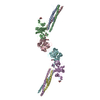
| ||||||||
|---|---|---|---|---|---|---|---|---|---|
| 1 |
| ||||||||
| Unit cell |
|
-Components
-Protein , 3 types, 6 molecules ADBECF
| #1: Protein | Mass: 14074.052 Da / Num. of mol.: 2 / Source method: isolated from a natural source / Source: (natural) #2: Protein | Mass: 37061.211 Da / Num. of mol.: 2 / Source method: isolated from a natural source / Source: (natural) #3: Protein | Mass: 38167.184 Da / Num. of mol.: 2 / Source method: isolated from a natural source / Source: (natural) |
|---|
-Protein/peptide / Sugars / Non-polymers , 3 types, 12 molecules GHIJ

| #4: Protein/peptide | Mass: 467.522 Da / Num. of mol.: 4 / Source method: obtained synthetically / Details: Synthetic peptide that occurs naturally in humans. / References: UniProt: P02675*PLUS #5: Sugar | ChemComp-NAG /  Type: D-saccharide, beta linking / Mass: 221.208 Da / Num. of mol.: 4 Type: D-saccharide, beta linking / Mass: 221.208 Da / Num. of mol.: 4Source method: isolated from a genetically manipulated source Formula: C8H15NO6 #6: Chemical | ChemComp-CA /  Mass: 40.078 Da / Num. of mol.: 4 / Source method: obtained synthetically / Formula: Ca Mass: 40.078 Da / Num. of mol.: 4 / Source method: obtained synthetically / Formula: Ca |
|---|
-Details
| Has protein modification | Y |
|---|
-Experimental details
-Experiment
| Experiment | Method: X-RAY DIFFRACTION / Number of used crystals: 1 |
|---|
- Sample preparation
| Crystal | Density Matthews: 3.18 Å3/Da / Density % sol: 61.35 % | |||||||||||||||||||||||||||||||||||||||||||||||||
|---|---|---|---|---|---|---|---|---|---|---|---|---|---|---|---|---|---|---|---|---|---|---|---|---|---|---|---|---|---|---|---|---|---|---|---|---|---|---|---|---|---|---|---|---|---|---|---|---|---|---|
| Crystal grow | Temperature: 295 K / Method: vapor diffusion, sitting drop / pH: 7 Details: PEG 3350 Tris buffer 5 mM CaCl2 2 mM GHRPam, pH 7.0, VAPOR DIFFUSION, SITTING DROP, temperature 295K | |||||||||||||||||||||||||||||||||||||||||||||||||
| Crystal grow | *PLUS | |||||||||||||||||||||||||||||||||||||||||||||||||
| Components of the solutions | *PLUS
|
-Data collection
| Diffraction | Mean temperature: 100 K |
|---|---|
| Diffraction source | Source: ROTATING ANODE / Type: RIGAKU / Wavelength: 1.54 Å |
| Detector | Type: MARRESEARCH / Detector: IMAGE PLATE / Date: Apr 23, 2002 |
| Radiation | Monochromator: OSMIC MIRRORS / Protocol: SINGLE WAVELENGTH / Monochromatic (M) / Laue (L): M / Scattering type: x-ray |
| Radiation wavelength | Wavelength: 1.54 Å / Relative weight: 1 |
| Reflection | Resolution: 2.9→20 Å / Num. all: 92116 / Num. obs: 75259 / % possible obs: 81.7 % / Observed criterion σ(F): 0 / Observed criterion σ(I): 0 / Rmerge(I) obs: 0.084 |
| Reflection shell | Resolution: 2.9→3 Å / % possible all: 66.7 |
| Reflection | *PLUS Num. obs: 40004 / Num. measured all: 75259 |
| Reflection shell | *PLUS % possible obs: 66.7 % |
- Processing
| Software |
| |||||||||||||||||||||||||
|---|---|---|---|---|---|---|---|---|---|---|---|---|---|---|---|---|---|---|---|---|---|---|---|---|---|---|
| Refinement | Method to determine structure: MOLECULAR REPLACEMENT / Resolution: 2.9→20 Å / σ(F): 0 / Stereochemistry target values: Engh & Huber
| |||||||||||||||||||||||||
| Refinement step | Cycle: LAST / Resolution: 2.9→20 Å
| |||||||||||||||||||||||||
| Refine LS restraints |
| |||||||||||||||||||||||||
| Refinement | *PLUS Highest resolution: 2.9 Å / Lowest resolution: 15 Å / % reflection Rfree: 5 % / Rfactor Rfree: 0.308 | |||||||||||||||||||||||||
| Solvent computation | *PLUS | |||||||||||||||||||||||||
| Displacement parameters | *PLUS |
