 Movie
Movie Controller


+ Open data

- Basic information
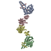
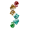
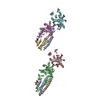
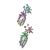
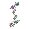
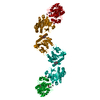
| Entry | Database: PDB / ID: 1n8e | ||||||
|---|---|---|---|---|---|---|---|
| Title | Fragment Double-D from Human Fibrin | ||||||
 Components Components |
| ||||||
 Keywords Keywords | BLOOD CLOTTING / Cross-linked fibrin / D:D interfaces / crystal packing | ||||||
| Function / homology |  Function and homology information Function and homology informationblood coagulation, common pathway / induction of bacterial agglutination / fibrinogen complex / Regulation of TLR by endogenous ligand / platelet alpha granule / cellular response to leptin stimulus / MyD88 deficiency (TLR2/4) / positive regulation of heterotypic cell-cell adhesion / IRAK4 deficiency (TLR2/4) / plasminogen activation ...blood coagulation, common pathway / induction of bacterial agglutination / fibrinogen complex / Regulation of TLR by endogenous ligand / platelet alpha granule / cellular response to leptin stimulus / MyD88 deficiency (TLR2/4) / positive regulation of heterotypic cell-cell adhesion / IRAK4 deficiency (TLR2/4) / plasminogen activation / MyD88:MAL(TIRAP) cascade initiated on plasma membrane / extracellular matrix structural constituent / p130Cas linkage to MAPK signaling for integrins / positive regulation of peptide hormone secretion / GRB2:SOS provides linkage to MAPK signaling for Integrins / positive regulation of exocytosis / blood coagulation, fibrin clot formation / protein polymerization / protein secretion / positive regulation of vasoconstriction / cellular response to interleukin-1 / Integrin cell surface interactions / : / negative regulation of endothelial cell apoptotic process / negative regulation of extrinsic apoptotic signaling pathway via death domain receptors / fibrinolysis / Integrin signaling / positive regulation of substrate adhesion-dependent cell spreading / cell adhesion molecule binding / platelet alpha granule lumen / cell-matrix adhesion / positive regulation of protein secretion / Post-translational protein phosphorylation / Signaling by high-kinase activity BRAF mutants / MAP2K and MAPK activation / response to calcium ion / platelet aggregation / Regulation of Insulin-like Growth Factor (IGF) transport and uptake by Insulin-like Growth Factor Binding Proteins (IGFBPs) / Signaling by RAF1 mutants / Signaling by moderate kinase activity BRAF mutants / Paradoxical activation of RAF signaling by kinase inactive BRAF / Signaling downstream of RAS mutants / Signaling by BRAF and RAF1 fusions / Platelet degranulation / extracellular vesicle / protein-folding chaperone binding / extracellular matrix / ER-Phagosome pathway / protein-containing complex assembly / blood microparticle / cell cortex / adaptive immune response / protein-macromolecule adaptor activity / positive regulation of ERK1 and ERK2 cascade / endoplasmic reticulum lumen / Amyloid fiber formation / signaling receptor binding / external side of plasma membrane / innate immune response / synapse / structural molecule activity / cell surface / endoplasmic reticulum / : / extracellular exosome / extracellular region / metal ion binding / plasma membrane Similarity search - Function | ||||||
| Biological species |  Homo sapiens (human) Homo sapiens (human) | ||||||
| Method |  X-RAY DIFFRACTION / SYNCHROTRON / MOLECULAR REPLACEMENT / Resolution: 4.5 Å X-RAY DIFFRACTION / SYNCHROTRON / MOLECULAR REPLACEMENT / Resolution: 4.5 Å | ||||||
 Authors Authors | Yang, Z. / Pandi, L. / Doolittle, R.F. | ||||||
 Citation Citation | Journal: Biochemistry / Year: 2002 Title: The Crystal Structure of Fragment Double-D from Cross-Linked Lamprey Fibrin Reveals Isopeptide Linkages across an Unexpected D-D Interface Authors: Yang, Z. / Pandi, L. / Doolittle, R.F. #1: Journal: Biochemistry / Year: 1998Title: Crystal Structure of Fragment Double-D from Human Fibrin with Two Different Bound Ligands Authors: Everse, S.J. / Spraggon, G. / Veerapandian, L. / Riley, M. / Doolittle, R.F. | ||||||
| History |
|
- Structure visualization
| Structure viewer | Molecule: MolmilJmol/JSmol |
|---|
- Downloads & links
-Download
| PDBx/mmCIF format | 1n8e.cif.gz | 55.9 KB | Display | PDBx/mmCIF format |
|---|---|---|---|---|
| PDB format | pdb1n8e.ent.gz | 31.1 KB | Display | PDB format |
| PDBx/mmJSON format | 1n8e.json.gz | Tree view | PDBx/mmJSON format | |
| Others |  Other downloads Other downloads |
-Validation report
| Arichive directory | https://data.pdbj.org/pub/pdb/validation_reports/n8/1n8eftp://data.pdbj.org/pub/pdb/validation_reports/n8/1n8e | HTTPS FTP |
|---|
-Related structure data
| Related structure data | 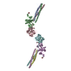 1n73C  1n86C  1fzcS S: Starting model for refinement C: citing same article ( |
|---|---|
| Similar structure data |


-Links
 PDBj
PDBj


- Assembly
| Deposited unit | 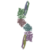
| ||||||||
|---|---|---|---|---|---|---|---|---|---|
| 1 |
| ||||||||
| Unit cell |
|
-Components
| #1: Protein | Mass: 10517.244 Da / Num. of mol.: 2 / Fragment: double-D alpha chain / Source method: isolated from a natural source / Details: Fibrinogen prepared from blood bank plasma / Source: (natural) Homo sapiens (human) / References: UniProt: P02671#2: Protein | Mass: 37691.992 Da / Num. of mol.: 2 / Fragment: double-D beta chain / Source method: isolated from a natural source / Details: Fibrinogen prepared from blood bank plasma / Source: (natural) Homo sapiens (human) / References: UniProt: P02675#3: Protein | Mass: 36693.754 Da / Num. of mol.: 2 / Fragment: double-D gamma chain / Source method: isolated from a natural source / Details: Fibrinogen prepared from blood bank plasma / Source: (natural) Homo sapiens (human) / References: UniProt: P02679 |
|---|
-Experimental details
-Experiment
| Experiment | Method: X-RAY DIFFRACTION / Number of used crystals: 1 |
|---|
- Sample preparation
| Crystal | Density Matthews: 2.88 Å3/Da / Density % sol: 57.27 % | |||||||||||||||||||||||||||||||||||||||||||||||||
|---|---|---|---|---|---|---|---|---|---|---|---|---|---|---|---|---|---|---|---|---|---|---|---|---|---|---|---|---|---|---|---|---|---|---|---|---|---|---|---|---|---|---|---|---|---|---|---|---|---|---|
| Crystal grow | Temperature: 295 K / Method: vapor diffusion, sitting drop / pH: 7.5 Details: PEG-3350, 50 mM Tris, 7.5 mM CaCl2, VAPOR DIFFUSION, SITTING DROP, temperature 295K | |||||||||||||||||||||||||||||||||||||||||||||||||
| Crystal grow | *PLUS pH: 7 | |||||||||||||||||||||||||||||||||||||||||||||||||
| Components of the solutions | *PLUS
|
-Data collection
| Diffraction | Mean temperature: 100 K |
|---|---|
| Diffraction source | Source: SYNCHROTRON / Site: SSRL  / Beamline: BL9-1 / Wavelength: 0.98 Å / Beamline: BL9-1 / Wavelength: 0.98 Å |
| Detector | Type: MARRESEARCH / Detector: IMAGE PLATE / Date: Feb 2, 1999 |
| Radiation | Monochromator: wiggler / Protocol: SINGLE WAVELENGTH / Monochromatic (M) / Laue (L): M / Scattering type: x-ray |
| Radiation wavelength | Wavelength: 0.98 Å / Relative weight: 1 |
| Reflection | Resolution: 4.5→30 Å / Num. all: 12247 / Num. obs: 10214 / % possible obs: 83.4 % / Observed criterion σ(F): 0 / Observed criterion σ(I): 0 / Rmerge(I) obs: 0.138 |
| Reflection shell | Resolution: 4.5→4.6 Å / % possible all: 53.3 |
| Reflection | *PLUS Num. measured all: 71273 |
| Reflection shell | *PLUS % possible obs: 53.3 % |
- Processing
| Software |
| ||||||||||||||||||||
|---|---|---|---|---|---|---|---|---|---|---|---|---|---|---|---|---|---|---|---|---|---|
| Refinement | Method to determine structure: MOLECULAR REPLACEMENT Starting model: fragment D of 1FZC Resolution: 4.5→30 Å / σ(F): 0 / σ(I): 0 / Stereochemistry target values: Engh & Huber / Details: The coordinates contain C-Alphas only.
| ||||||||||||||||||||
| Refinement step | Cycle: LAST / Resolution: 4.5→30 Å
| ||||||||||||||||||||
| Software | *PLUS Name: CNS / Version: 1 / Classification: refinement | ||||||||||||||||||||
| Refinement | *PLUS Lowest resolution: 20 Å / % reflection Rfree: 5 % | ||||||||||||||||||||
| Solvent computation | *PLUS | ||||||||||||||||||||
| Displacement parameters | *PLUS |
