 Movie
Movie Controller

[English] 日本語
 Yorodumi
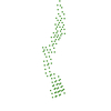
- PDB-1lwu: Crystal structure of fragment D from lamprey fibrinogen complexed... -
+ Open data

- Basic information
| Entry | Database: PDB / ID: 1lwu | ||||||||||||
|---|---|---|---|---|---|---|---|---|---|---|---|---|---|
| Title | Crystal structure of fragment D from lamprey fibrinogen complexed with the peptide Gly-His-Arg-Pro-amide | ||||||||||||
 Components Components |
| ||||||||||||
 Keywords Keywords | BLOOD CLOTTING / heterotrimer / protein-peptide complex | ||||||||||||
| Function / homology |  Function and homology information Function and homology informationblood coagulation, common pathway / fibrinogen complex / positive regulation of heterotypic cell-cell adhesion / extracellular matrix structural constituent / protein polymerization / fibrinolysis / platelet aggregation / protein-macromolecule adaptor activity / signaling receptor binding / metal ion binding Similarity search - Function | ||||||||||||
| Biological species |  | ||||||||||||
| Method |  X-RAY DIFFRACTION / SYNCHROTRON / MOLECULAR REPLACEMENT / Resolution: 2.8 Å X-RAY DIFFRACTION / SYNCHROTRON / MOLECULAR REPLACEMENT / Resolution: 2.8 Å | ||||||||||||
 Authors Authors | Yang, Z. / Spraggon, G. / Pandi, L. / Everse, S.J. / Riley, M. / Doolittle, R.F. | ||||||||||||
 Citation Citation | Journal: Biochemistry / Year: 2002 Title: Crystal structure of fragment D from lamprey fibrinogen complexed with the peptide Gly-His-Arg-Pro-amide. Authors: Yang, Z. / Spraggon, G. / Pandi, L. / Everse, S.J. / Riley, M. / Doolittle, R.F. | ||||||||||||
| History |
|
- Structure visualization
| Structure viewer | Molecule: MolmilJmol/JSmol |
|---|
- Downloads & links
-Download
| PDBx/mmCIF format | 1lwu.cif.gz | 607.9 KB | Display | PDBx/mmCIF format |
|---|---|---|---|---|
| PDB format | pdb1lwu.ent.gz | 498.9 KB | Display | PDB format |
| PDBx/mmJSON format | 1lwu.json.gz | Tree view | PDBx/mmJSON format | |
| Others |  Other downloads Other downloads |
-Validation report
| Arichive directory | https://data.pdbj.org/pub/pdb/validation_reports/lw/1lwuftp://data.pdbj.org/pub/pdb/validation_reports/lw/1lwu | HTTPS FTP |
|---|
-Related structure data
| Related structure data | |
|---|---|
| Similar structure data |
-Links
 PDBj
PDBj


- Assembly
| Deposited unit | 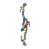
| ||||||||
|---|---|---|---|---|---|---|---|---|---|
| 1 | 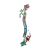
| ||||||||
| 2 | 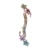
| ||||||||
| Unit cell |
|
-Components
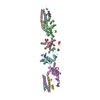
-Protein , 3 types, 12 molecules ADGJBEHKCFIL
| #1: Protein | Mass: 14074.052 Da / Num. of mol.: 4 / Fragment: fragment / Source method: isolated from a natural source / Source: (natural) #2: Protein | Mass: 37061.211 Da / Num. of mol.: 4 / Fragment: Segment 2 of 2 / Source method: isolated from a natural source / Source: (natural) #3: Protein | Mass: 37475.555 Da / Num. of mol.: 4 / Source method: isolated from a natural source / Source: (natural) |
|---|
-Protein/peptide / Non-polymers , 2 types, 12 molecules MNOP
| #4: Protein/peptide | Mass: 465.530 Da / Num. of mol.: 4 / Source method: obtained synthetically #8: Chemical | ChemComp-CA /  Mass: 40.078 Da / Num. of mol.: 8 / Source method: obtained synthetically / Formula: Ca Mass: 40.078 Da / Num. of mol.: 8 / Source method: obtained synthetically / Formula: Ca |
|---|
-Sugars , 6 types, 30 molecules 


| #5: Polysaccharide | 2-acetamido-2-deoxy-beta-D-glucopyranose-(1-4)-2-acetamido-2-deoxy-beta-D-glucopyranose Source method: isolated from a genetically manipulated source | ||||||||
|---|---|---|---|---|---|---|---|---|---|
| #6: Sugar | ChemComp-NDG /  Type: D-saccharide, alpha linking / Mass: 221.208 Da / Num. of mol.: 9 Type: D-saccharide, alpha linking / Mass: 221.208 Da / Num. of mol.: 9Source method: isolated from a genetically manipulated source Formula: C8H15NO6 #7: Sugar | ChemComp-MAN /  Type: D-saccharide, alpha linking / Mass: 180.156 Da / Num. of mol.: 10 Type: D-saccharide, alpha linking / Mass: 180.156 Da / Num. of mol.: 10Source method: isolated from a genetically manipulated source Formula: C6H12O6 #9: Sugar | ChemComp-NAG /  Type: D-saccharide, beta linking / Mass: 221.208 Da / Num. of mol.: 7 Type: D-saccharide, beta linking / Mass: 221.208 Da / Num. of mol.: 7Source method: isolated from a genetically manipulated source Formula: C8H15NO6 #10: Sugar |  Type: D-saccharide, beta linking / Mass: 180.156 Da / Num. of mol.: 2 Type: D-saccharide, beta linking / Mass: 180.156 Da / Num. of mol.: 2Source method: isolated from a genetically manipulated source Formula: C6H12O6 #11: Sugar | ChemComp-GAL / |  Type: D-saccharide, beta linking / Mass: 180.156 Da / Num. of mol.: 1 Type: D-saccharide, beta linking / Mass: 180.156 Da / Num. of mol.: 1Source method: isolated from a genetically manipulated source Formula: C6H12O6 |
-Details
| Has protein modification | Y |
|---|
-Experimental details
-Experiment
| Experiment | Method: X-RAY DIFFRACTION / Number of used crystals: 1 |
|---|
- Sample preparation
| Crystal | Density Matthews: 2.48 Å3/Da / Density % sol: 50.47 % | ||||||||||||||||||||||||||||||||||||||||||||||||||||||||
|---|---|---|---|---|---|---|---|---|---|---|---|---|---|---|---|---|---|---|---|---|---|---|---|---|---|---|---|---|---|---|---|---|---|---|---|---|---|---|---|---|---|---|---|---|---|---|---|---|---|---|---|---|---|---|---|---|---|
| Crystal grow | Temperature: 295 K / Method: vapor diffusion, sitting drop / pH: 6.75 Details: PEG 3350, MES buffer, pH 6.75, 10 mM CaCl2, VAPOR DIFFUSION, SITTING DROP at 295K | ||||||||||||||||||||||||||||||||||||||||||||||||||||||||
| Crystal grow | *PLUS pH: 7 | ||||||||||||||||||||||||||||||||||||||||||||||||||||||||
| Components of the solutions | *PLUS
|
-Data collection
| Diffraction | Mean temperature: 110 K |
|---|---|
| Diffraction source | Source: SYNCHROTRON / Site: NSLS  / Beamline: X12C / Wavelength: 1.1 Å / Beamline: X12C / Wavelength: 1.1 Å |
| Detector | Type: BRANDEIS - B4 / Detector: CCD / Date: Aug 22, 1998 |
| Radiation | Monochromator: Si 111 CHANNEL / Protocol: SINGLE WAVELENGTH / Monochromatic (M) / Laue (L): M / Scattering type: x-ray |
| Radiation wavelength | Wavelength: 1.1 Å / Relative weight: 1 |
| Reflection | Resolution: 2.8→20 Å / Num. all: 74102 / Num. obs: 74102 / % possible obs: 88.3 % / Observed criterion σ(F): 0 / Observed criterion σ(I): 0 / Rmerge(I) obs: 0.078 |
| Reflection shell | Resolution: 2.8→2.87 Å / % possible all: 58.4 |
| Reflection | *PLUS Num. measured all: 200629 |
| Reflection shell | *PLUS % possible obs: 58.4 % |
- Processing
| Software |
| |||||||||||||||||||||||||
|---|---|---|---|---|---|---|---|---|---|---|---|---|---|---|---|---|---|---|---|---|---|---|---|---|---|---|
| Refinement | Method to determine structure: MOLECULAR REPLACEMENT / Resolution: 2.8→20 Å / Isotropic thermal model: Isotropic / Cross valid method: THROUGHOUT / σ(F): 0 / Stereochemistry target values: Engh & Huber
| |||||||||||||||||||||||||
| Refinement step | Cycle: LAST / Resolution: 2.8→20 Å
| |||||||||||||||||||||||||
| Refine LS restraints |
| |||||||||||||||||||||||||
| Refinement | *PLUS Highest resolution: 2.8 Å / % reflection Rfree: 5 % / Rfactor Rfree: 0.289 / Rfactor Rwork: 0.241 | |||||||||||||||||||||||||
| Solvent computation | *PLUS | |||||||||||||||||||||||||
| Displacement parameters | *PLUS |
