 Movie
Movie Controller

[English] 日本語
 Yorodumi
- PDB-1mrs: CRYSTAL STRUCTURE OF MYCOBACTERIUM TUBERCULOSIS THYMIDYLATE KINAS... -
+ Open data

- Basic information
| Entry | Database: PDB / ID: 1mrs | ||||||
|---|---|---|---|---|---|---|---|
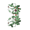
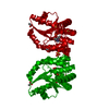
| Title | CRYSTAL STRUCTURE OF MYCOBACTERIUM TUBERCULOSIS THYMIDYLATE KINASE COMPLEXED WITH 5-CH2OH DEOXYURIDINE MONOPHOSPHATE | ||||||
 Components Components | THYMIDYLATE KINASE | ||||||
 Keywords Keywords | TRANSFERASE / TRANSFERASE (ATP:TMP PHOSPHOTRANSFERASE) / KINASE | ||||||
| Function / homology |  Function and homology information Function and homology informationTMP metabolic process / dTMP kinase / dUDP biosynthetic process / dTDP biosynthetic process / dTMP kinase activity / dTTP biosynthetic process / GTP binding / magnesium ion binding / protein homodimerization activity / ATP binding ...TMP metabolic process / dTMP kinase / dUDP biosynthetic process / dTDP biosynthetic process / dTMP kinase activity / dTTP biosynthetic process / GTP binding / magnesium ion binding / protein homodimerization activity / ATP binding / metal ion binding / cytosol / cytoplasm Similarity search - Function | ||||||
| Biological species |   Mycobacterium tuberculosis (bacteria) Mycobacterium tuberculosis (bacteria) | ||||||
| Method |  X-RAY DIFFRACTION / SYNCHROTRON / MOLECULAR REPLACEMENT / Resolution: 2 Å X-RAY DIFFRACTION / SYNCHROTRON / MOLECULAR REPLACEMENT / Resolution: 2 Å | ||||||
 Authors Authors | Haouz, A. / Vanheusden, V. / Munier-Lehmann, H. / Froeyen, M. / Herdewijn, P. / Van Calenbergh, S. / Delarue, M. | ||||||
 Citation Citation | Journal: J.Biol.Chem. / Year: 2003 Title: Enzymatic and structural analysis of inhibitors designed against Mycobacterium tuberculosis thymidylate kinase. New insights into the phosphoryl transfer mechanism. Authors: Haouz, A. / Vanheusden, V. / Munier-Lehmann, H. / Froeyen, M. | ||||||
| History |
|
- Structure visualization
| Structure viewer | Molecule: MolmilJmol/JSmol |
|---|
- Downloads & links
-Download
| PDBx/mmCIF format | 1mrs.cif.gz | 55.1 KB | Display | PDBx/mmCIF format |
|---|---|---|---|---|
| PDB format | pdb1mrs.ent.gz | 38.4 KB | Display | PDB format |
| PDBx/mmJSON format | 1mrs.json.gz | Tree view | PDBx/mmJSON format | |
| Others |  Other downloads Other downloads |
-Validation report
| Arichive directory | https://data.pdbj.org/pub/pdb/validation_reports/mr/1mrsftp://data.pdbj.org/pub/pdb/validation_reports/mr/1mrs | HTTPS FTP |
|---|
-Related structure data
| Related structure data | 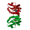 1mrnC 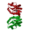 1g3uS S: Starting model for refinement C: citing same article ( |
|---|---|
| Similar structure data |

-Links
 PDBj
PDBj- Assembly
| Deposited unit | 
| ||||||||
|---|---|---|---|---|---|---|---|---|---|
| 1 | 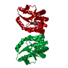
| ||||||||
| Unit cell |
| ||||||||
| Details | The molecule is a dimer in solution. Monomer B can be generated from monomer A through the transformation X, -Y, -Z |
-Components
| #1: Protein | Mass: 22662.525 Da / Num. of mol.: 1 Source method: isolated from a genetically manipulated source Source: (gene. exp.) Mycobacterium tuberculosis (bacteria) / Plasmid: PET22B / Production host: References: UniProt: O05891, UniProt: P9WKE1*PLUS, dTMP kinase | ||||||
|---|---|---|---|---|---|---|---|
| #2: Chemical |   Mass: 96.063 Da / Num. of mol.: 2 / Source method: obtained synthetically / Formula: SO4 Mass: 96.063 Da / Num. of mol.: 2 / Source method: obtained synthetically / Formula: SO4#3: Chemical | ChemComp-MG / |   Mass: 24.305 Da / Num. of mol.: 1 / Source method: obtained synthetically / Formula: Mg Mass: 24.305 Da / Num. of mol.: 1 / Source method: obtained synthetically / Formula: Mg#4: Chemical | ChemComp-5HU / |   Type: DNA linking / Mass: 338.208 Da / Num. of mol.: 1 / Source method: obtained synthetically / Formula: C10H15N2O9P Type: DNA linking / Mass: 338.208 Da / Num. of mol.: 1 / Source method: obtained synthetically / Formula: C10H15N2O9P#5: Water | ChemComp-HOH / |  Mass: 18.015 Da / Num. of mol.: 77 / Source method: isolated from a natural source / Formula: H2O Mass: 18.015 Da / Num. of mol.: 77 / Source method: isolated from a natural source / Formula: H2O |
-Experimental details
-Experiment
| Experiment | Method: X-RAY DIFFRACTION / Number of used crystals: 1 |
|---|
- Sample preparation
| Crystal | Density Matthews: 2.5 Å3/Da / Density % sol: 50.76 % | ||||||||||||||||||||||||||||||||||||||||||||||||
|---|---|---|---|---|---|---|---|---|---|---|---|---|---|---|---|---|---|---|---|---|---|---|---|---|---|---|---|---|---|---|---|---|---|---|---|---|---|---|---|---|---|---|---|---|---|---|---|---|---|
| Crystal grow | Temperature: 290 K / Method: vapor diffusion, hanging drop / pH: 6 Details: Ammonium sulfate, PEG600, MES, magnesium acetate, pH 6.0, VAPOR DIFFUSION, HANGING DROP, temperature 290K | ||||||||||||||||||||||||||||||||||||||||||||||||
| Crystal grow | *PLUS Method: vapor diffusion | ||||||||||||||||||||||||||||||||||||||||||||||||
| Components of the solutions | *PLUS
|
-Data collection
| Diffraction | Mean temperature: 110 K |
|---|---|
| Diffraction source | Source: SYNCHROTRON / Site: ESRF  / Beamline: ID14-1 / Wavelength: 0.934 Å / Beamline: ID14-1 / Wavelength: 0.934 Å |
| Detector | Type: ADSC QUANTUM 4 / Detector: CCD / Date: Jun 10, 2001 |
| Radiation | Monochromator: Si 111 / Protocol: SINGLE WAVELENGTH / Monochromatic (M) / Laue (L): M / Scattering type: x-ray |
| Radiation wavelength | Wavelength: 0.934 Å / Relative weight: 1 |
| Reflection | Resolution: 2→25 Å / Num. all: 14739 / Num. obs: 14739 / % possible obs: 89.5 % / Observed criterion σ(F): 0 / Observed criterion σ(I): 0 / Redundancy: 6 % / Rmerge(I) obs: 0.082 / Rsym value: 0.082 / Net I/σ(I): 27.8 |
| Reflection shell | Resolution: 2→2.07 Å / Redundancy: 5.9 % / Rmerge(I) obs: 0.271 / Mean I/σ(I) obs: 18.8 / Rsym value: 0.271 / % possible all: 98.6 |
| Reflection | *PLUS |
| Reflection shell | *PLUS % possible obs: 98.6 % / Num. unique obs: 1575 |
- Processing
| Software |
| |||||||||||||||||||||||||
|---|---|---|---|---|---|---|---|---|---|---|---|---|---|---|---|---|---|---|---|---|---|---|---|---|---|---|
| Refinement | Method to determine structure: MOLECULAR REPLACEMENT Starting model: PDB ENTRY 1G3U Resolution: 2→25 Å / Isotropic thermal model: isotropic / Cross valid method: THROUGHOUT / σ(F): 1 / Stereochemistry target values: Engh & Huber
| |||||||||||||||||||||||||
| Displacement parameters | Biso mean: 36.1318 Å2 | |||||||||||||||||||||||||
| Refine analyze |
| |||||||||||||||||||||||||
| Refinement step | Cycle: LAST / Resolution: 2→25 Å
| |||||||||||||||||||||||||
| Refine LS restraints |
| |||||||||||||||||||||||||
| LS refinement shell | Resolution: 2→2.07 Å
| |||||||||||||||||||||||||
| Refinement | *PLUS % reflection Rfree: 5 % | |||||||||||||||||||||||||
| Solvent computation | *PLUS | |||||||||||||||||||||||||
| Displacement parameters | *PLUS | |||||||||||||||||||||||||
| Refine LS restraints | *PLUS
|
