 Movie
Movie Controller

[English] 日本語
 Yorodumi
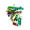
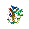
- PDB-1mqk: Crystal structure of the unliganded Fv-fragment of the anti-cytoc... -
+ Open data

- Basic information
| Entry | Database: PDB / ID: 1mqk | ||||||
|---|---|---|---|---|---|---|---|
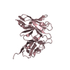
| Title | Crystal structure of the unliganded Fv-fragment of the anti-cytochrome C oxidase antibody 7E2 | ||||||
 Components Components |
| ||||||
 Keywords Keywords | IMMUNE SYSTEM / antibody / membrane protein / cytochrome C oxidase / high-resolution structure | ||||||
| Function / homology |  Function and homology information Function and homology informationimmunoglobulin mediated immune response / immunoglobulin complex / antigen binding / adaptive immune response / immune response / extracellular region / plasma membrane Similarity search - Function | ||||||
| Biological species |  | ||||||
| Method |  X-RAY DIFFRACTION / SYNCHROTRON / MOLECULAR REPLACEMENT / Resolution: 1.28 Å X-RAY DIFFRACTION / SYNCHROTRON / MOLECULAR REPLACEMENT / Resolution: 1.28 Å | ||||||
 Authors Authors | Essen, L.-O. / Harrenga, A. / Ostermeier, C. / Michel, H. | ||||||
 Citation Citation | Journal: Acta Crystallogr.,Sect.D / Year: 2003 Title: 1.3 A X-ray structure of an antibody Fv fragment used for induced membrane-protein crystallization. Authors: Essen, L.O. / Harrenga, A. / Ostermeier, C. / Michel, H. #1: Journal: Proteins / Year: 1995Title: Crystals of an antibody Fv fragment against an integral membrane protein diffracting to 1.28 resolution Authors: Ostermeier, C. / Essen, L.-O. / Michel, H. #2: Journal: Nat.Struct.Biol. / Year: 1995Title: Fv fragment-mediated crystallization of the membrane protein bacterial cytochrome C oxidase Authors: Ostermeier, C. / Iwata, S. / Ludwig, B. / Michel, H. #3: Journal: Nature / Year: 1995Title: Structure at 2.8 resolution of cytochrome C oxidase from Paracoccus denitrificans Authors: Iwata, S. / Ostermeier, C. / Ludwig, B. / Michel, H. #4: Journal: J.Biol.Chem. / Year: 1999Title: The cytochrome C oxidase from Paracoccus denitrificans does not change the metal center upon reduction Authors: Harrenga, A. / Michel, H. #5: Journal: Proc.Natl.Acad.Sci.USA / Year: 1997Title: Structure at 2.7 resolution of the Paracoccus denitrificans two-subunit cytochrome C oxidase complexed with an antibody Fv fragment Authors: Ostermeier, C. / Harrenga, A. / Ermler, U. / Michel, H. | ||||||
| History |
| ||||||
| Remark 999 | SEQUENCE ACCORDING TO THE AUTHOR, THE SEQUENCE FOR THE HEAVY AND LIGHT CHAINS OF ANTIBODY 7E2 HAS ...SEQUENCE ACCORDING TO THE AUTHOR, THE SEQUENCE FOR THE HEAVY AND LIGHT CHAINS OF ANTIBODY 7E2 HAS NOT BEEN DEPOSITED IN ANY SEQUENCE DATABASE. SOME SEQUENCE INFORMATION IS PRESENT IN PDB ENTRY 1QLE. |
- Structure visualization
| Structure viewer | Molecule: MolmilJmol/JSmol |
|---|
- Downloads & links
-Download
| PDBx/mmCIF format | 1mqk.cif.gz | 68.5 KB | Display | PDBx/mmCIF format |
|---|---|---|---|---|
| PDB format | pdb1mqk.ent.gz | 49.6 KB | Display | PDB format |
| PDBx/mmJSON format | 1mqk.json.gz | Tree view | PDBx/mmJSON format | |
| Others |  Other downloads Other downloads |
-Validation report
| Arichive directory | https://data.pdbj.org/pub/pdb/validation_reports/mq/1mqkftp://data.pdbj.org/pub/pdb/validation_reports/mq/1mqk | HTTPS FTP |
|---|
-Related structure data
| Related structure data |  1qleS S: Starting model for refinement |
|---|---|
| Similar structure data |


-Links
 PDBj
PDBj


- Assembly
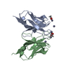
| Deposited unit | 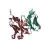
| ||||||||
|---|---|---|---|---|---|---|---|---|---|
| 1 |
| ||||||||
| Unit cell |
|
-Components
| #1: Antibody | Mass: 13260.795 Da / Num. of mol.: 1 Source method: isolated from a genetically manipulated source Source: (gene. exp.)  |
|---|---|
| #2: Antibody | Mass: 14324.923 Da / Num. of mol.: 1 Source method: isolated from a genetically manipulated source Source: (gene. exp.) |
| #3: Water | ChemComp-HOH /  Mass: 18.015 Da / Num. of mol.: 318 / Source method: isolated from a natural source / Formula: H2O Mass: 18.015 Da / Num. of mol.: 318 / Source method: isolated from a natural source / Formula: H2O |
| Has protein modification | Y |
-Experimental details
-Experiment
| Experiment | Method: X-RAY DIFFRACTION / Number of used crystals: 1 |
|---|
- Sample preparation
| Crystal | Density Matthews: 2.61 Å3/Da / Density % sol: 52.83 % | ||||||||||||||||||
|---|---|---|---|---|---|---|---|---|---|---|---|---|---|---|---|---|---|---|---|
| Crystal grow | Temperature: 277 K / Method: microdialysis / pH: 6 Details: sodium phosphate, pH 6.0, MICRODIALYSIS, temperature 277K | ||||||||||||||||||
| Crystal | *PLUS Density % sol: 53 % | ||||||||||||||||||
| Crystal grow | *PLUS | ||||||||||||||||||
| Components of the solutions | *PLUS
|
-Data collection
| Diffraction | Mean temperature: 277 K |
|---|---|
| Diffraction source | Source: SYNCHROTRON / Site: MPG/DESY, HAMBURG  / Beamline: BW6 / Wavelength: 1 Å / Beamline: BW6 / Wavelength: 1 Å |
| Detector | Type: MARRESEARCH / Detector: IMAGE PLATE / Date: Jun 12, 1993 / Details: mirrors |
| Radiation | Monochromator: mirrors / Protocol: SINGLE WAVELENGTH / Monochromatic (M) / Laue (L): M / Scattering type: x-ray |
| Radiation wavelength | Wavelength: 1 Å / Relative weight: 1 |
| Reflection | Resolution: 1.28→22.5 Å / Num. obs: 64251 / % possible obs: 85.1 % / Observed criterion σ(F): 0 / Observed criterion σ(I): 1 / Redundancy: 2.8 % / Biso Wilson estimate: 13.2 Å2 / Rmerge(I) obs: 0.4 / Net I/σ(I): 28.6 |
| Reflection shell | Resolution: 1.28→1.31 Å / Redundancy: 1.9 % / Rmerge(I) obs: 0.218 / Mean I/σ(I) obs: 5.2 / % possible all: 63.4 |
| Reflection | *PLUS Num. measured all: 178789 / Rmerge(I) obs: 0.04 |
| Reflection shell | *PLUS % possible obs: 63.4 % |
- Processing
| Software |
| ||||||||||||||||||||||||
|---|---|---|---|---|---|---|---|---|---|---|---|---|---|---|---|---|---|---|---|---|---|---|---|---|---|
| Refinement | Method to determine structure: MOLECULAR REPLACEMENT Starting model: PDB ENTRY 1QLE Resolution: 1.28→8 Å / Isotropic thermal model: anisotropic / σ(F): 0 / Stereochemistry target values: Engh & Huber Details: Used weighted full matrix least squares procedure of SHELX-93 to generate model. Only isotropic model provided
| ||||||||||||||||||||||||
| Displacement parameters | Biso mean: 22.8 Å2 | ||||||||||||||||||||||||
| Refine analyze | Luzzati coordinate error obs: 0.1 Å / Luzzati sigma a obs: 0.07 Å | ||||||||||||||||||||||||
| Refinement step | Cycle: LAST / Resolution: 1.28→8 Å
| ||||||||||||||||||||||||
| Refine LS restraints |
| ||||||||||||||||||||||||
| Software | *PLUS Name: SHELXL / Version: 93 / Classification: refinement | ||||||||||||||||||||||||
| Refinement | *PLUS Lowest resolution: 8 Å / % reflection Rfree: 4 % | ||||||||||||||||||||||||
| Solvent computation | *PLUS | ||||||||||||||||||||||||
| Displacement parameters | *PLUS | ||||||||||||||||||||||||
| Refine LS restraints | *PLUS
|
