 Movie
Movie Controller

[English] 日本語
 Yorodumi
- PDB-1ar1: Structure at 2.7 Angstrom Resolution of the Paracoccus Denitrific... -
+ Open data

- Basic information
| Entry | Database: PDB / ID: 1ar1 | ||||||
|---|---|---|---|---|---|---|---|
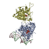
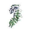
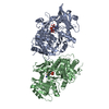
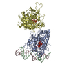
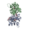
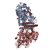
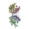
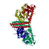
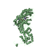
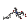
| Title | Structure at 2.7 Angstrom Resolution of the Paracoccus Denitrificans two-subunit Cytochrome C Oxidase Complexed with an Antibody Fv Fragment | ||||||
 Components Components |
| ||||||
 Keywords Keywords | COMPLEX (OXIDOREDUCTASE/ANTIBODY) / COMPLEX (OXIDOREDUCTASE-ANTIBODY) / ELECTRON TRANSPORT / TRANSMEMBRANE / CYTOCHROME OXIDASE / ANTIBODY COMPLEX / COMPLEX (OXIDOREDUCTASE-ANTIBODY) complex | ||||||
| Function / homology |  Function and homology information Function and homology informationrespiratory chain complex IV / cytochrome-c oxidase / oxidative phosphorylation / cytochrome-c oxidase activity / immunoglobulin mediated immune response / electron transport coupled proton transport / immunoglobulin complex / antigen binding / ATP synthesis coupled electron transport / respiratory electron transport chain ...respiratory chain complex IV / cytochrome-c oxidase / oxidative phosphorylation / cytochrome-c oxidase activity / immunoglobulin mediated immune response / electron transport coupled proton transport / immunoglobulin complex / antigen binding / ATP synthesis coupled electron transport / respiratory electron transport chain / adaptive immune response / oxidoreductase activity / immune response / copper ion binding / heme binding / extracellular region / metal ion binding / plasma membrane Similarity search - Function | ||||||
| Biological species |  Paracoccus denitrificans (bacteria) Paracoccus denitrificans (bacteria) | ||||||
| Method |  X-RAY DIFFRACTION / SYNCHROTRON / MOLECULAR REPLACEMENT / Resolution: 2.7 Å X-RAY DIFFRACTION / SYNCHROTRON / MOLECULAR REPLACEMENT / Resolution: 2.7 Å | ||||||
 Authors Authors | Ostermeier, C. / Harrenga, A. / Ermler, U. / Michel, H. | ||||||
 Citation Citation | Journal: Proc.Natl.Acad.Sci.USA / Year: 1997 Title: Structure at 2.7 A resolution of the Paracoccus denitrificans two-subunit cytochrome c oxidase complexed with an antibody FV fragment. Authors: Ostermeier, C. / Harrenga, A. / Ermler, U. / Michel, H. | ||||||
| History |
|
- Structure visualization
| Structure viewer | Molecule: MolmilJmol/JSmol |
|---|
- Downloads & links
-Download
| PDBx/mmCIF format | 1ar1.cif.gz | 220.7 KB | Display | PDBx/mmCIF format |
|---|---|---|---|---|
| PDB format | pdb1ar1.ent.gz | 173 KB | Display | PDB format |
| PDBx/mmJSON format | 1ar1.json.gz | Tree view | PDBx/mmJSON format | |
| Others |  Other downloads Other downloads |
-Validation report
| Arichive directory | https://data.pdbj.org/pub/pdb/validation_reports/ar/1ar1ftp://data.pdbj.org/pub/pdb/validation_reports/ar/1ar1 | HTTPS FTP |
|---|
-Related structure data
| Similar structure data |
|---|

-Links
 PDBj
PDBj


- Assembly
| Deposited unit | 
| ||||||||
|---|---|---|---|---|---|---|---|---|---|
| 1 |
| ||||||||
| Unit cell |
|
-Components
-CYTOCHROME C ... , 2 types, 2 molecules AB
| #1: Protein | Mass: 62486.645 Da / Num. of mol.: 1 Source method: isolated from a genetically manipulated source Source: (gene. exp.) Paracoccus denitrificans (bacteria) / Cellular location: CYTOPLASMIC MEMBRANE / Production host: |
|---|---|
| #2: Protein | Mass: 32563.643 Da / Num. of mol.: 1 Source method: isolated from a genetically manipulated source Source: (gene. exp.) Paracoccus denitrificans (bacteria) / Cellular location: CYTOPLASMIC MEMBRANE / Production host: |
-Antibody , 2 types, 2 molecules CD
| #3: Antibody | Mass: 14324.923 Da / Num. of mol.: 1 Source method: isolated from a genetically manipulated source Source: (gene. exp.) |
|---|---|
| #4: Antibody | Mass: 13260.795 Da / Num. of mol.: 1 Source method: isolated from a genetically manipulated source Source: (gene. exp.) |
-Non-polymers , 6 types, 68 molecules 


| #5: Chemical |  Mass: 63.546 Da / Num. of mol.: 3 / Source method: obtained synthetically / Formula: Cu Mass: 63.546 Da / Num. of mol.: 3 / Source method: obtained synthetically / Formula: Cu#6: Chemical | ChemComp-MG / |  Mass: 24.305 Da / Num. of mol.: 1 / Source method: obtained synthetically / Formula: Mg Mass: 24.305 Da / Num. of mol.: 1 / Source method: obtained synthetically / Formula: Mg#7: Chemical | ChemComp-CA / |  Mass: 40.078 Da / Num. of mol.: 1 / Source method: obtained synthetically / Formula: Ca Mass: 40.078 Da / Num. of mol.: 1 / Source method: obtained synthetically / Formula: Ca#8: Chemical |  Mass: 852.837 Da / Num. of mol.: 2 / Source method: obtained synthetically / Formula: C49H56FeN4O6 Mass: 852.837 Da / Num. of mol.: 2 / Source method: obtained synthetically / Formula: C49H56FeN4O6#9: Chemical | ChemComp-LDA /  Mass: 229.402 Da / Num. of mol.: 9 / Source method: obtained synthetically / Formula: C14H31NO / Comment: LDAO, detergent*YM Mass: 229.402 Da / Num. of mol.: 9 / Source method: obtained synthetically / Formula: C14H31NO / Comment: LDAO, detergent*YM#10: Water | ChemComp-HOH / | Mass: 18.015 Da / Num. of mol.: 52 / Source method: isolated from a natural source / Formula: H2O |
|---|
-Details
| Has protein modification | Y |
|---|
-Experimental details
-Experiment
| Experiment | Method: X-RAY DIFFRACTION / Number of used crystals: 10 |
|---|
- Sample preparation
| Crystal | Density Matthews: 4.8 Å3/Da / Density % sol: 72 % | |||||||||||||||
|---|---|---|---|---|---|---|---|---|---|---|---|---|---|---|---|---|
| Crystal grow | pH: 5.5 / Details: pH 5.5 | |||||||||||||||
| Crystal grow | *PLUS Temperature: 293 K / Method: vapor diffusion, hanging drop | |||||||||||||||
| Components of the solutions | *PLUS
|
-Data collection
| Diffraction | Mean temperature: 277 K |
|---|---|
| Diffraction source | Source: SYNCHROTRON / Site: ESRF  / Beamline: BM14 / Wavelength: 1.078 / Beamline: BM14 / Wavelength: 1.078 |
| Detector | Type: PRINCETON 2K / Detector: CCD / Date: Jan 31, 1997 / Details: COLLIMATING AND FOCUSSING MIRROR |
| Radiation | Monochromator: SI(111) / Monochromatic (M) / Laue (L): M / Scattering type: x-ray |
| Radiation wavelength | Wavelength: 1.078 Å / Relative weight: 1 |
| Reflection | Resolution: 2.7→50 Å / Num. obs: 57373 / % possible obs: 93.8 % / Observed criterion σ(I): 0 / Redundancy: 3.4 % / Biso Wilson estimate: 44.2 Å2 / Rmerge(I) obs: 0.068 / Net I/σ(I): 13.4 |
| Reflection shell | Resolution: 2.7→2.8 Å / Redundancy: 2.5 % / Rmerge(I) obs: 0.332 / Mean I/σ(I) obs: 4 / % possible all: 79.7 |
| Reflection | *PLUS Num. obs: 57945 / Num. measured all: 198657 |
- Processing
| Software |
| ||||||||||||||||||||||||||||||||||||||||||||||||||||||||||||||||||||||||||||||||
|---|---|---|---|---|---|---|---|---|---|---|---|---|---|---|---|---|---|---|---|---|---|---|---|---|---|---|---|---|---|---|---|---|---|---|---|---|---|---|---|---|---|---|---|---|---|---|---|---|---|---|---|---|---|---|---|---|---|---|---|---|---|---|---|---|---|---|---|---|---|---|---|---|---|---|---|---|---|---|---|---|---|
| Refinement | Method to determine structure: MOLECULAR REPLACEMENT Starting model: FOUR PROTEIN SUBUNITS CONTAINING CYTOCHROME C OXIDASE FROM PARACOCCUS DENITRIFICANS (NATURE 376: 660-669) Resolution: 2.7→30 Å / Rfactor Rfree error: 0.005 / Data cutoff high absF: 10000000 / Data cutoff low absF: 0.001 / Isotropic thermal model: RESTRAINED / Cross valid method: THROUGHOUT / σ(F): 2
| ||||||||||||||||||||||||||||||||||||||||||||||||||||||||||||||||||||||||||||||||
| Displacement parameters | Biso mean: 59.7 Å2 | ||||||||||||||||||||||||||||||||||||||||||||||||||||||||||||||||||||||||||||||||
| Refine analyze |
| ||||||||||||||||||||||||||||||||||||||||||||||||||||||||||||||||||||||||||||||||
| Refinement step | Cycle: LAST / Resolution: 2.7→30 Å
| ||||||||||||||||||||||||||||||||||||||||||||||||||||||||||||||||||||||||||||||||
| Refine LS restraints |
| ||||||||||||||||||||||||||||||||||||||||||||||||||||||||||||||||||||||||||||||||
| LS refinement shell | Resolution: 2.7→2.87 Å / Rfactor Rfree error: 0.018 / Total num. of bins used: 6
| ||||||||||||||||||||||||||||||||||||||||||||||||||||||||||||||||||||||||||||||||
| Xplor file |
| ||||||||||||||||||||||||||||||||||||||||||||||||||||||||||||||||||||||||||||||||
| Software | *PLUS Name: X-PLOR / Version: 3.851 / Classification: refinement | ||||||||||||||||||||||||||||||||||||||||||||||||||||||||||||||||||||||||||||||||
| Refine LS restraints | *PLUS
|
