 Movie
Movie Controller

[English] 日本語
 Yorodumi
- PDB-1ktr: Crystal Structure of the Anti-His Tag Antibody 3D5 Single-Chain F... -
+ Open data

- Basic information
| Entry | Database: PDB / ID: 1ktr | |||||||||
|---|---|---|---|---|---|---|---|---|---|---|
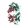
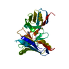
| Title | Crystal Structure of the Anti-His Tag Antibody 3D5 Single-Chain Fragment (scFv) in Complex with a Oligohistidine peptide | |||||||||
 Components Components |
| |||||||||
 Keywords Keywords | IMMUNE SYSTEM / immunoglobulin domains / single chain antibody-antigen complex / His tag recognition / scfv | |||||||||
| Function / homology | Immunoglobulins / Immunoglobulin-like / Sandwich / Mainly Beta Function and homology information Function and homology information | |||||||||
| Biological species |  synthetic construct (others) | |||||||||
| Method |  X-RAY DIFFRACTION / SYNCHROTRON / MOLECULAR REPLACEMENT / Resolution: 2.7 Å X-RAY DIFFRACTION / SYNCHROTRON / MOLECULAR REPLACEMENT / Resolution: 2.7 Å | |||||||||
 Authors Authors | Kaufmann, M. / Lindner, P. / Honegger, A. / Blank, K. / Tschopp, M. / Capitani, G. / Plueckthun, A. / Gruetter, M.G. | |||||||||
 Citation Citation | Journal: J.Mol.Biol. / Year: 2002 Title: Crystal structure of the anti-His tag antibody 3D5 single-chain fragment complexed to its antigen. Authors: Kaufmann, M. / Lindner, P. / Honegger, A. / Blank, K. / Tschopp, M. / Capitani, G. / Pluckthun, A. / Grutter, M.G. #1: Journal: BIO*TECHNIQUES / Year: 1997Title: Specific detection of his-tagged proteins with recombinant anti-His tag scFv-phosphatase or scFv-phage fusions Authors: Lindner, P. / Bauer, K. / Krebber, A. / Nieba, L. / Kremmer, E. / Krebber, C. / Honegger, A. / Klinger, B. / Mocikat, R. / Plueckthun, A. | |||||||||
| History |
| |||||||||
| Remark 999 | SEQUENCE Neither the sequence of the wild-type nor the mutated sequence, from which the structure ...SEQUENCE Neither the sequence of the wild-type nor the mutated sequence, from which the structure was solved, was deposited to a database. In comparison to the wild-type of the anti-his tag antibody 3d5 variable light chain, the mutated sequence chain L has the following mutations: L9S, V78F, Y88D. In comparison to the wild-type of the anti-his tag antibody 3d5 variable heavy chain, the mutated sequence chain H has the following mutations: L12D, H48P, S51G, K77R, E100D, L144T. The scfv, consisting of chains L, M, and H, was expressed as one polypeptide. The N-terminal variable light chain, chain L and the C-terminal variable heavy chain, chain H are connected by the peptide linker, chain M. Note that chains L, M, and H are one continuous polypeptide chain and most of the linker, chain M, is invisible. The first three residues of chain L, DYK, are part of the flag recognition tag. |
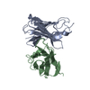
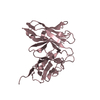
- Structure visualization
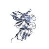
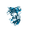
| Structure viewer | Molecule: MolmilJmol/JSmol |
|---|
- Downloads & links
-Download
| PDBx/mmCIF format | 1ktr.cif.gz | 63.6 KB | Display | PDBx/mmCIF format |
|---|---|---|---|---|
| PDB format | pdb1ktr.ent.gz | 44.9 KB | Display | PDB format |
| PDBx/mmJSON format | 1ktr.json.gz | Tree view | PDBx/mmJSON format | |
| Others |  Other downloads Other downloads |
-Validation report
| Arichive directory | https://data.pdbj.org/pub/pdb/validation_reports/kt/1ktrftp://data.pdbj.org/pub/pdb/validation_reports/kt/1ktr | HTTPS FTP |
|---|
-Related structure data


-Links
 PDBj
PDBj

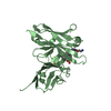
- Assembly
| Deposited unit | 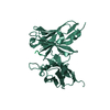
| ||||||||
|---|---|---|---|---|---|---|---|---|---|
| 1 |
| ||||||||
| Unit cell |
|
-Components
| #1: Antibody | Mass: 26475.104 Da / Num. of mol.: 1 Mutation: L9S, V78F, Y88D, L12D, H48P, S51G, K77R, E100D, L144T Source method: isolated from a genetically manipulated source Source: (gene. exp.)  |
|---|---|
| #2: Protein/peptide | Mass: 846.896 Da / Num. of mol.: 1 / Source method: obtained synthetically / Details: The peptide was chemically synthetized. / Source: (synth.) synthetic construct (others) |
| #3: Water | ChemComp-HOH /  Mass: 18.015 Da / Num. of mol.: 156 / Source method: isolated from a natural source / Formula: H2O Mass: 18.015 Da / Num. of mol.: 156 / Source method: isolated from a natural source / Formula: H2O |
| Compound details | The peptide linker connects the variable light chain and the variable heavy chain. |
| Has protein modification | Y |
| Sequence details | Neither the sequence of the wild-type nor the mutated sequence, from which the structure was ...Neither the sequence of the wild-type nor the mutated sequence, from which the structure was solved, was deposited to a database. In comparison to the wild-type of the anti-his tag antibody 3d5 variable light chain, the mutated sequence chain L has the following mutations: L9S, V78F, Y88D. In comparison to the wild-type of the anti-his tag antibody 3d5 variable heavy chain, the mutated sequence chain H has the following mutations: L12D, H48P, S51G, K77R, E100D, L144T. The first three residues DYK, are part of the flag recognition tag. |
-Experimental details
-Experiment
| Experiment | Method: X-RAY DIFFRACTION / Number of used crystals: 1 |
|---|
- Sample preparation
| Crystal | Density Matthews: 5.4 Å3/Da / Density % sol: 77 % | ||||||||||||||||||||||||||||||||||||||||||
|---|---|---|---|---|---|---|---|---|---|---|---|---|---|---|---|---|---|---|---|---|---|---|---|---|---|---|---|---|---|---|---|---|---|---|---|---|---|---|---|---|---|---|---|
| Crystal grow | Temperature: 277 K / Method: vapor diffusion, sitting drop / pH: 6.4 Details: PEG 8000, magnesium acetate, MES, pH 6.4, VAPOR DIFFUSION, SITTING DROP, temperature 277K | ||||||||||||||||||||||||||||||||||||||||||
| Crystal grow | *PLUS Temperature: 4 ℃ | ||||||||||||||||||||||||||||||||||||||||||
| Components of the solutions | *PLUS
|
-Data collection
| Diffraction | Mean temperature: 100 K |
|---|---|
| Diffraction source | Source: SYNCHROTRON / Site: ESRF  / Beamline: BM1A / Wavelength: 0.873 Å / Beamline: BM1A / Wavelength: 0.873 Å |
| Detector | Type: MAR scanner 345 mm plate / Detector: IMAGE PLATE / Date: Oct 2, 2000 / Details: Mirrors |
| Radiation | Monochromator: MIRRORS / Protocol: SINGLE WAVELENGTH / Monochromatic (M) / Laue (L): M / Scattering type: x-ray |
| Radiation wavelength | Wavelength: 0.873 Å / Relative weight: 1 |
| Reflection | Resolution: 2.7→30 Å / Num. obs: 16996 / % possible obs: 99 % / Observed criterion σ(I): -2.5 / Redundancy: 7.54 % / Biso Wilson estimate: 52.8 Å2 / Rmerge(I) obs: 0.116 / Net I/σ(I): 16.6 |
| Reflection shell | Resolution: 2.7→2.8 Å / Rmerge(I) obs: 0.537 / Mean I/σ(I) obs: 4.4 / % possible all: 99.8 |
| Reflection | *PLUS Lowest resolution: 30 Å / Redundancy: 7.6 % / Num. measured all: 128480 |
- Processing
| Software |
| |||||||||||||||||||||||||
|---|---|---|---|---|---|---|---|---|---|---|---|---|---|---|---|---|---|---|---|---|---|---|---|---|---|---|
| Refinement | Method to determine structure: MOLECULAR REPLACEMENT Starting model: VL: PDB ENTRY 1TET, VH: PDB ENTRY 1PSK Resolution: 2.7→30 Å / Isotropic thermal model: Isotropic / Cross valid method: THROUGHOUT / σ(F): 0 / Stereochemistry target values: Engh & Huber
| |||||||||||||||||||||||||
| Displacement parameters | Biso mean: 33.4 Å2
| |||||||||||||||||||||||||
| Refine analyze |
| |||||||||||||||||||||||||
| Refinement step | Cycle: LAST / Resolution: 2.7→30 Å
| |||||||||||||||||||||||||
| Refine LS restraints |
| |||||||||||||||||||||||||
| LS refinement shell | Resolution: 2.7→2.87 Å / Rfactor Rfree error: 0.024
| |||||||||||||||||||||||||
| Refinement | *PLUS Lowest resolution: 30 Å / Rfactor obs: 0.1904 / Rfactor Rfree: 0.222 / Rfactor Rwork: 0.1904 | |||||||||||||||||||||||||
| Solvent computation | *PLUS | |||||||||||||||||||||||||
| Displacement parameters | *PLUS | |||||||||||||||||||||||||
| Refine LS restraints | *PLUS
| |||||||||||||||||||||||||
| LS refinement shell | *PLUS Highest resolution: 2.7 Å / Rfactor obs: 0.261 |
