 Movie
Movie Controller


+ Open data

- Basic information
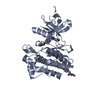
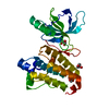
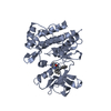
| Entry | Database: PDB / ID: 1mpu | ||||||
|---|---|---|---|---|---|---|---|
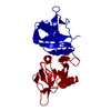
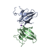
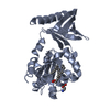
| Title | Crystal Structure of the free human NKG2D immunoreceptor | ||||||
 Components Components | NKG2-D type II integral membrane protein | ||||||
 Keywords Keywords | IMMUNE SYSTEM / C-type lectin-like domain | ||||||
| Function / homology |  Function and homology information Function and homology informationnegative regulation of natural killer cell chemotaxis / MHC class Ib receptor activity / negative regulation of GTPase activity / positive regulation of natural killer cell mediated cytotoxicity / natural killer cell activation / natural killer cell mediated cytotoxicity / stimulatory C-type lectin receptor signaling pathway / MHC class I protein binding / T cell costimulation / nitric oxide biosynthetic process ...negative regulation of natural killer cell chemotaxis / MHC class Ib receptor activity / negative regulation of GTPase activity / positive regulation of natural killer cell mediated cytotoxicity / natural killer cell activation / natural killer cell mediated cytotoxicity / stimulatory C-type lectin receptor signaling pathway / MHC class I protein binding / T cell costimulation / nitric oxide biosynthetic process / DAP12 interactions / Immunoregulatory interactions between a Lymphoid and a non-Lymphoid cell / positive regulation of nitric oxide biosynthetic process / positive regulation of type II interferon production / DAP12 signaling / cellular response to lipopolysaccharide / carbohydrate binding / signaling receptor activity / adaptive immune response / cell differentiation / defense response to Gram-positive bacterium / external side of plasma membrane / cell surface / signal transduction / membrane / identical protein binding / plasma membrane Similarity search - Function | ||||||
| Biological species |  Homo sapiens (human) Homo sapiens (human) | ||||||
| Method |  X-RAY DIFFRACTION / MOLECULAR REPLACEMENT / Resolution: 2.5 Å X-RAY DIFFRACTION / MOLECULAR REPLACEMENT / Resolution: 2.5 Å | ||||||
 Authors Authors | McFarland, B.J. / Kortemme, T. / Baker, D. / Strong, R.K. | ||||||
 Citation Citation | Journal: Structure / Year: 2003 Title: Symmetry Recognizing Asymmetry: Analysis of the Interactions between the C-Type Lectin-like Immunoreceptor NKG2D and MHC Class I-like Ligands Authors: McFarland, B.J. / Kortemme, T. / Yu, S.F. / Baker, D. / Strong, R.K. | ||||||
| History |
|
- Structure visualization
| Structure viewer | Molecule: MolmilJmol/JSmol |
|---|
- Downloads & links
-Download
| PDBx/mmCIF format | 1mpu.cif.gz | 42.6 KB | Display | PDBx/mmCIF format |
|---|---|---|---|---|
| PDB format | pdb1mpu.ent.gz | 29.4 KB | Display | PDB format |
| PDBx/mmJSON format | 1mpu.json.gz | Tree view | PDBx/mmJSON format | |
| Others |  Other downloads Other downloads |
-Validation report
| Arichive directory | https://data.pdbj.org/pub/pdb/validation_reports/mp/1mpuftp://data.pdbj.org/pub/pdb/validation_reports/mp/1mpu | HTTPS FTP |
|---|
-Related structure data
| Related structure data |  1hyrS S: Starting model for refinement |
|---|---|
| Similar structure data |


-Links
 PDBj
PDBj


- Assembly
| Deposited unit | 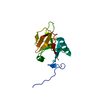
| ||||||||
|---|---|---|---|---|---|---|---|---|---|
| 1 | 
| ||||||||
| Unit cell |
| ||||||||
| Details | The biological assembly is a homodimer is generated by the two-fold axis: 1/2+x, 1/2-y, 3/4-z. |
-Components
| #1: Protein | Mass: 15981.102 Da / Num. of mol.: 1 / Fragment: residues 80-216 Source method: isolated from a genetically manipulated source Source: (gene. exp.) Homo sapiens (human) / Plasmid: pET22b+ / Species (production host): Escherichia coli / Production host:  |
|---|---|
| #2: Chemical | ChemComp-PO4 /   Mass: 94.971 Da / Num. of mol.: 1 / Source method: obtained synthetically / Formula: PO4 Mass: 94.971 Da / Num. of mol.: 1 / Source method: obtained synthetically / Formula: PO4 |
| #3: Water | ChemComp-HOH /  Mass: 18.015 Da / Num. of mol.: 105 / Source method: isolated from a natural source / Formula: H2O Mass: 18.015 Da / Num. of mol.: 105 / Source method: isolated from a natural source / Formula: H2O |
| Has protein modification | Y |
-Experimental details
-Experiment
| Experiment | Method: X-RAY DIFFRACTION / Number of used crystals: 1 |
|---|
- Sample preparation
| Crystal | Density Matthews: 2.17 Å3/Da / Density % sol: 43.32 % | ||||||||||||||||||||||||||||||||||||||||||||||||||||||||||||||||||||||
|---|---|---|---|---|---|---|---|---|---|---|---|---|---|---|---|---|---|---|---|---|---|---|---|---|---|---|---|---|---|---|---|---|---|---|---|---|---|---|---|---|---|---|---|---|---|---|---|---|---|---|---|---|---|---|---|---|---|---|---|---|---|---|---|---|---|---|---|---|---|---|---|
| Crystal grow | Temperature: 291 K / Method: vapor diffusion, hanging drop / pH: 9 Details: ammonium dihydrogen phosphate, bicine, ethylene glycol, pH 9.0, VAPOR DIFFUSION, HANGING DROP, temperature 291K | ||||||||||||||||||||||||||||||||||||||||||||||||||||||||||||||||||||||
| Crystal grow | *PLUS pH: 7 / Method: vapor diffusion, sitting drop | ||||||||||||||||||||||||||||||||||||||||||||||||||||||||||||||||||||||
| Components of the solutions | *PLUS
|
-Data collection
| Diffraction | Mean temperature: 200 K |
|---|---|
| Diffraction source | Source: ROTATING ANODE / Type: RIGAKU RU200 / Wavelength: 1.5418 Å |
| Detector | Type: RIGAKU RAXIS IV / Detector: IMAGE PLATE / Date: Dec 5, 2001 / Details: Osmics mirrors |
| Radiation | Protocol: SINGLE WAVELENGTH / Monochromatic (M) / Laue (L): M / Scattering type: x-ray |
| Radiation wavelength | Wavelength: 1.5418 Å / Relative weight: 1 |
| Reflection | Resolution: 2.5→20 Å / Num. all: 5240 / Num. obs: 5057 / % possible obs: 96.5 % / Observed criterion σ(F): 0 / Observed criterion σ(I): 0 / Redundancy: 21.1 % / Rsym value: 0.066 / Net I/σ(I): 20.2 |
| Reflection shell | Resolution: 2.5→2.59 Å / Mean I/σ(I) obs: 6.8 / Num. unique all: 517 / Rsym value: 0.199 / % possible all: 90.1 |
| Reflection | *PLUS Highest resolution: 2.5 Å / Rmerge(I) obs: 0.066 |
| Reflection shell | *PLUS % possible obs: 90.1 % / Num. unique obs: 466 / Rmerge(I) obs: 0.199 |
- Processing
| Software |
| |||||||||||||||||||||||||
|---|---|---|---|---|---|---|---|---|---|---|---|---|---|---|---|---|---|---|---|---|---|---|---|---|---|---|
| Refinement | Method to determine structure: MOLECULAR REPLACEMENT Starting model: PDB ENTRY 1HYR (CHAIN A) Resolution: 2.5→20 Å / Isotropic thermal model: anisotropic / Cross valid method: THROUGHOUT / σ(F): 0 / Stereochemistry target values: Engh & Huber
| |||||||||||||||||||||||||
| Displacement parameters |
| |||||||||||||||||||||||||
| Refine analyze | Luzzati coordinate error obs: 0.37 Å / Luzzati d res low obs: 5 Å / Luzzati sigma a obs: 0.5 Å | |||||||||||||||||||||||||
| Refinement step | Cycle: LAST / Resolution: 2.5→20 Å
| |||||||||||||||||||||||||
| Refine LS restraints |
| |||||||||||||||||||||||||
| LS refinement shell | Resolution: 2.5→2.59 Å
| |||||||||||||||||||||||||
| Refinement | *PLUS Highest resolution: 2.5 Å / Lowest resolution: 20 Å | |||||||||||||||||||||||||
| Solvent computation | *PLUS | |||||||||||||||||||||||||
| Displacement parameters | *PLUS | |||||||||||||||||||||||||
| Refine LS restraints | *PLUS
| |||||||||||||||||||||||||
| LS refinement shell | *PLUS Highest resolution: 2.5 Å |
