 Movie
Movie Controller

[English] 日本語
 Yorodumi
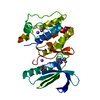
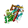
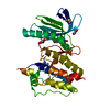
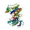
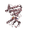
- PDB-1ozu: Crystal Structure of Familial ALS Mutant S134N of human Cu,Zn Sup... -
+ Open data

- Basic information
| Entry | Database: PDB / ID: 1ozu | ||||||
|---|---|---|---|---|---|---|---|
| Title | Crystal Structure of Familial ALS Mutant S134N of human Cu,Zn Superoxide Dismutase (CuZnSOD) to 1.3A resolution | ||||||
 Components Components | Superoxide dismutase [Cu-Zn] | ||||||
 Keywords Keywords | OXIDOREDUCTASE / beta barrel / amyloid-like linear filaments | ||||||
| Function / homology |  Function and homology information Function and homology informationaction potential initiation / response to antipsychotic drug / neurofilament cytoskeleton organization / response to carbon monoxide / relaxation of vascular associated smooth muscle / regulation of organ growth / dense core granule / response to superoxide / positive regulation of oxidative stress-induced intrinsic apoptotic signaling pathway / peripheral nervous system myelin maintenance ...action potential initiation / response to antipsychotic drug / neurofilament cytoskeleton organization / response to carbon monoxide / relaxation of vascular associated smooth muscle / regulation of organ growth / dense core granule / response to superoxide / positive regulation of oxidative stress-induced intrinsic apoptotic signaling pathway / peripheral nervous system myelin maintenance / anterograde axonal transport / protein phosphatase 2B binding / regulation of T cell differentiation in thymus / Oxidoreductases; Acting on a sulfur group of donors / regulation of GTPase activity / retina homeostasis / auditory receptor cell stereocilium organization / cellular response to potassium ion / hydrogen peroxide biosynthetic process / retrograde axonal transport / myeloid cell homeostasis / superoxide anion generation / superoxide metabolic process / muscle cell cellular homeostasis / response to copper ion / superoxide dismutase / Detoxification of Reactive Oxygen Species / superoxide dismutase activity / heart contraction / cellular response to cadmium ion / cellular response to ATP / negative regulation of reproductive process / negative regulation of developmental process / transmission of nerve impulse / regulation of multicellular organism growth / ectopic germ cell programmed cell death / response to axon injury / ovarian follicle development / neuronal action potential / positive regulation of superoxide anion generation / axon cytoplasm / removal of superoxide radicals / embryo implantation / reactive oxygen species metabolic process / Gene and protein expression by JAK-STAT signaling after Interleukin-12 stimulation / positive regulation of phagocytosis / dendrite cytoplasm / placenta development / thymus development / positive regulation of cytokine production / determination of adult lifespan / response to amphetamine / regulation of mitochondrial membrane potential / glutathione metabolic process / response to hydrogen peroxide / locomotory behavior / sensory perception of sound / response to nutrient levels / mitochondrial intermembrane space / negative regulation of inflammatory response / regulation of blood pressure / small GTPase binding / Platelet degranulation / peroxisome / response to heat / protein-folding chaperone binding / cytoplasmic vesicle / spermatogenesis / gene expression / intracellular iron ion homeostasis / response to ethanol / negative regulation of neuron apoptotic process / positive regulation of MAPK cascade / lysosome / positive regulation of apoptotic process / mitochondrial matrix / response to xenobiotic stimulus / copper ion binding / neuronal cell body / apoptotic process / protein homodimerization activity / protein-containing complex / mitochondrion / : / extracellular exosome / extracellular region / zinc ion binding / nucleoplasm / identical protein binding / nucleus / cytoplasm / cytosol Similarity search - Function | ||||||
| Biological species |  Homo sapiens (human) Homo sapiens (human) | ||||||
| Method |  X-RAY DIFFRACTION / SYNCHROTRON / MOLECULAR REPLACEMENT / Resolution: 1.3 Å X-RAY DIFFRACTION / SYNCHROTRON / MOLECULAR REPLACEMENT / Resolution: 1.3 Å | ||||||
 Authors Authors | Elam, J.S. / Taylor, A.B. / Strange, R. / Antonyuk, S. / Doucette, P.A. / Rodriguez, J.A. / Hasnain, S.S. / Hayward, L.J. / Valentine, J.S. / Yeates, T.O. / Hart, P.J. | ||||||
 Citation Citation | Journal: Nat.Struct.Biol. / Year: 2003 Title: Amyloid-like Filaments and Water-filled Nanotubes Formed by SOD1 Mutant Proteins Linked to Familial ALS Authors: Elam, J.S. / Taylor, A.B. / Strange, R. / Antonyuk, S. / Doucette, P.A. / Rodriguez, J.A. / Hasnain, S.S. / Hayward, L.J. / Valentine, J.S. / Yeates, T.O. / Hart, P.J. | ||||||
| History |
|
- Structure visualization
| Structure viewer | Molecule: MolmilJmol/JSmol |
|---|
- Downloads & links
-Download
| PDBx/mmCIF format | 1ozu.cif.gz | 120.3 KB | Display | PDBx/mmCIF format |
|---|---|---|---|---|
| PDB format | pdb1ozu.ent.gz | 93.1 KB | Display | PDB format |
| PDBx/mmJSON format | 1ozu.json.gz | Tree view | PDBx/mmJSON format | |
| Others |  Other downloads Other downloads |
-Validation report
| Arichive directory | https://data.pdbj.org/pub/pdb/validation_reports/oz/1ozuftp://data.pdbj.org/pub/pdb/validation_reports/oz/1ozu | HTTPS FTP |
|---|
-Related structure data
| Related structure data |  1oezC  1oztC  1azvS C: citing same article ( S: Starting model for refinement |
|---|---|
| Similar structure data |


-Links
 PDBj
PDBj


- Assembly
| Deposited unit | 
| ||||||||
|---|---|---|---|---|---|---|---|---|---|
| 1 |
| ||||||||
| Unit cell |
| ||||||||
| Details | The biological assembly is a homodimer. There is one homodimer in asymmetric unit. |
-Components
| #1: Protein | Mass: 15870.586 Da / Num. of mol.: 2 / Mutation: S134N Source method: isolated from a genetically manipulated source Source: (gene. exp.) Homo sapiens (human) / Gene: SOD1 / Plasmid: YEp 351 / Production host:  #2: Chemical |   Mass: 65.409 Da / Num. of mol.: 3 / Source method: obtained synthetically / Formula: Zn Mass: 65.409 Da / Num. of mol.: 3 / Source method: obtained synthetically / Formula: Zn#3: Chemical |   Mass: 96.063 Da / Num. of mol.: 2 / Source method: obtained synthetically / Formula: SO4 Mass: 96.063 Da / Num. of mol.: 2 / Source method: obtained synthetically / Formula: SO4#4: Water | ChemComp-HOH / |  Mass: 18.015 Da / Num. of mol.: 247 / Source method: isolated from a natural source / Formula: H2O Mass: 18.015 Da / Num. of mol.: 247 / Source method: isolated from a natural source / Formula: H2OHas protein modification | Y | |
|---|
-Experimental details
-Experiment
| Experiment | Method: X-RAY DIFFRACTION / Number of used crystals: 1 |
|---|
- Sample preparation
| Crystal | Density Matthews: 1.89 Å3/Da / Density % sol: 34.28 % | ||||||||||||||||||||||||||||||
|---|---|---|---|---|---|---|---|---|---|---|---|---|---|---|---|---|---|---|---|---|---|---|---|---|---|---|---|---|---|---|---|
| Crystal grow | Temperature: 298 K / Method: vapor diffusion, hanging drop / pH: 7 Details: Ammonium sulfate, pH 7.0, VAPOR DIFFUSION, HANGING DROP, temperature 298.0K | ||||||||||||||||||||||||||||||
| Crystal grow | *PLUS Temperature: 25 ℃ / pH: 6.5 / Method: vapor diffusion, hanging drop | ||||||||||||||||||||||||||||||
| Components of the solutions | *PLUS
|
-Data collection
| Diffraction | Mean temperature: 100 K |
|---|---|
| Diffraction source | Source: SYNCHROTRON / Site: NSLS  / Beamline: X8C / Wavelength: 1.1 Å / Beamline: X8C / Wavelength: 1.1 Å |
| Detector | Type: ADSC QUANTUM 4 / Detector: CCD / Date: Apr 21, 2002 / Details: mirrors |
| Radiation | Monochromator: mirrors / Protocol: SINGLE WAVELENGTH / Monochromatic (M) / Laue (L): M / Scattering type: x-ray |
| Radiation wavelength | Wavelength: 1.1 Å / Relative weight: 1 |
| Reflection | Resolution: 1.3→52.7 Å / Num. all: 58507 / Num. obs: 58507 / % possible obs: 97.9 % / Observed criterion σ(I): -3 / Redundancy: 4.62 % / Biso Wilson estimate: 12.6 Å2 / Rmerge(I) obs: 0.05 / Net I/σ(I): 23.7 |
| Reflection shell | Resolution: 1.3→1.38 Å / Rmerge(I) obs: 0.498 / Mean I/σ(I) obs: 2.86 / Num. unique all: 5121 / % possible all: 87.1 |
| Reflection | *PLUS Highest resolution: 1.3 Å / Lowest resolution: 50 Å / Num. measured all: 270095 / Rmerge(I) obs: 0.05 |
| Reflection shell | *PLUS % possible obs: 87.1 % |
- Processing
| Software |
| ||||||||||||||||||||||||||||||||||||||||||||||||||||||||||||||||||||||||||||||||||||||||||||||||||||||||||||||||||||||||||||||||||
|---|---|---|---|---|---|---|---|---|---|---|---|---|---|---|---|---|---|---|---|---|---|---|---|---|---|---|---|---|---|---|---|---|---|---|---|---|---|---|---|---|---|---|---|---|---|---|---|---|---|---|---|---|---|---|---|---|---|---|---|---|---|---|---|---|---|---|---|---|---|---|---|---|---|---|---|---|---|---|---|---|---|---|---|---|---|---|---|---|---|---|---|---|---|---|---|---|---|---|---|---|---|---|---|---|---|---|---|---|---|---|---|---|---|---|---|---|---|---|---|---|---|---|---|---|---|---|---|---|---|---|---|
| Refinement | Method to determine structure: MOLECULAR REPLACEMENT Starting model: PDB Entry 1AZV Resolution: 1.3→52.7 Å / Cor.coef. Fo:Fc: 0.964 / Cor.coef. Fo:Fc free: 0.961 / SU B: 2.158 / SU ML: 0.048 / Isotropic thermal model: Anisotropic / Cross valid method: THROUGHOUT / σ(F): 0 / σ(I): 0 / ESU R: 0.06 / ESU R Free: 0.052 / Stereochemistry target values: MAXIMUM LIKELIHOOD / Details: HYDROGENS HAVE BEEN ADDED IN THE RIDING POSITIONS
| ||||||||||||||||||||||||||||||||||||||||||||||||||||||||||||||||||||||||||||||||||||||||||||||||||||||||||||||||||||||||||||||||||
| Solvent computation | Ion probe radii: 0.8 Å / Shrinkage radii: 0.8 Å / VDW probe radii: 1.4 Å / Solvent model: BABINET MODEL WITH MASK | ||||||||||||||||||||||||||||||||||||||||||||||||||||||||||||||||||||||||||||||||||||||||||||||||||||||||||||||||||||||||||||||||||
| Displacement parameters | Biso mean: 13.417 Å2
| ||||||||||||||||||||||||||||||||||||||||||||||||||||||||||||||||||||||||||||||||||||||||||||||||||||||||||||||||||||||||||||||||||
| Refinement step | Cycle: LAST / Resolution: 1.3→52.7 Å
| ||||||||||||||||||||||||||||||||||||||||||||||||||||||||||||||||||||||||||||||||||||||||||||||||||||||||||||||||||||||||||||||||||
| Refine LS restraints |
| ||||||||||||||||||||||||||||||||||||||||||||||||||||||||||||||||||||||||||||||||||||||||||||||||||||||||||||||||||||||||||||||||||
| LS refinement shell | Resolution: 1.303→1.337 Å / Total num. of bins used: 20 /
| ||||||||||||||||||||||||||||||||||||||||||||||||||||||||||||||||||||||||||||||||||||||||||||||||||||||||||||||||||||||||||||||||||
| Refinement | *PLUS Highest resolution: 1.3 Å / Lowest resolution: 21.1 Å / Rfactor Rfree: 0.211 / Rfactor Rwork: 0.188 | ||||||||||||||||||||||||||||||||||||||||||||||||||||||||||||||||||||||||||||||||||||||||||||||||||||||||||||||||||||||||||||||||||
| Solvent computation | *PLUS | ||||||||||||||||||||||||||||||||||||||||||||||||||||||||||||||||||||||||||||||||||||||||||||||||||||||||||||||||||||||||||||||||||
| Displacement parameters | *PLUS | ||||||||||||||||||||||||||||||||||||||||||||||||||||||||||||||||||||||||||||||||||||||||||||||||||||||||||||||||||||||||||||||||||
| Refine LS restraints | *PLUS
|
