positive regulation of oxidative stress-induced intrinsic apoptotic signaling pathway / action potential initiation / regulation of T cell differentiation in thymus / response to antipsychotic drug / neurofilament cytoskeleton organization / response to carbon monoxide / regulation of organ growth / relaxation of vascular associated smooth muscle / peripheral nervous system myelin maintenance / response to superoxide ...positive regulation of oxidative stress-induced intrinsic apoptotic signaling pathway / action potential initiation / regulation of T cell differentiation in thymus / response to antipsychotic drug / neurofilament cytoskeleton organization / response to carbon monoxide / regulation of organ growth / relaxation of vascular associated smooth muscle / peripheral nervous system myelin maintenance / response to superoxide / anterograde axonal transport / protein phosphatase 2B binding / dense core granule / Oxidoreductases; Acting on a sulfur group of donors / regulation of GTPase activity / auditory receptor cell stereocilium organization / retina homeostasis / hydrogen peroxide biosynthetic process / cellular response to potassium ion / retrograde axonal transport / myeloid cell homeostasis / superoxide anion generation / muscle cell cellular homeostasis / superoxide metabolic process / response to copper ion / superoxide dismutase / Detoxification of Reactive Oxygen Species / heart contraction / superoxide dismutase activity / cellular response to cadmium ion / regulation of multicellular organism growth / cellular response to ATP / negative regulation of reproductive process / negative regulation of developmental process / transmission of nerve impulse / response to axon injury / ectopic germ cell programmed cell death / ovarian follicle development / neuronal action potential / embryo implantation / positive regulation of superoxide anion generation / axon cytoplasm / removal of superoxide radicals / reactive oxygen species metabolic process / thymus development / Gene and protein expression by JAK-STAT signaling after Interleukin-12 stimulation / placenta development / positive regulation of phagocytosis / dendrite cytoplasm / response to amphetamine / determination of adult lifespan / positive regulation of cytokine production / regulation of mitochondrial membrane potential / glutathione metabolic process / response to hydrogen peroxide / sensory perception of sound / locomotory behavior / response to nutrient levels / negative regulation of inflammatory response / mitochondrial intermembrane space / regulation of blood pressure / small GTPase binding / Platelet degranulation / peroxisome / response to heat / protein-folding chaperone binding / gene expression / cytoplasmic vesicle / spermatogenesis / negative regulation of neuron apoptotic process / intracellular iron ion homeostasis / response to ethanol / positive regulation of MAPK cascade / lysosome / response to xenobiotic stimulus / positive regulation of apoptotic process / mitochondrial matrix / copper ion binding / neuronal cell body / apoptotic process / protein homodimerization activity / protein-containing complex / mitochondrion / : / extracellular exosome / extracellular region / zinc ion binding / nucleoplasm / identical protein binding / nucleus / cytosol / cytoplasm Similarity search - Function
In the structure databanks used in Yorodumi, some data are registered as the other names, "COVID-19 virus" and "2019-nCoV". Here are the details of the virus and the list of structure data.
Jan 31, 2019. EMDB accession codes are about to change! (news from PDBe EMDB page)
EMDB accession codes are about to change! (news from PDBe EMDB page)
The allocation of 4 digits for EMDB accession codes will soon come to an end. Whilst these codes will remain in use, new EMDB accession codes will include an additional digit and will expand incrementally as the available range of codes is exhausted. The current 4-digit format prefixed with “EMD-” (i.e. EMD-XXXX) will advance to a 5-digit format (i.e. EMD-XXXXX), and so on. It is currently estimated that the 4-digit codes will be depleted around Spring 2019, at which point the 5-digit format will come into force.
The EM Navigator/Yorodumi systems omit the EMD- prefix.
Related info.:Q: What is EMD? / ID/Accession-code notation in Yorodumi/EM Navigator
Yorodumi is a browser for structure data from EMDB, PDB, SASBDB, etc.
This page is also the successor to EM Navigator detail page, and also detail information page/front-end page for Omokage search.
The word "yorodu" (or yorozu) is an old Japanese word meaning "ten thousand". "mi" (miru) is to see.
Related info.:EMDB / PDB / SASBDB / Comparison of 3 databanks / Yorodumi Search / Aug 31, 2016. New EM Navigator & Yorodumi / Yorodumi Papers / Jmol/JSmol / Function and homology information / Changes in new EM Navigator and Yorodumi
 Movie
Movie Controller


 Open data
 Basic information
 Components
 Keywords
 Function and homology information
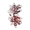
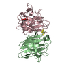
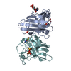
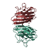
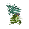
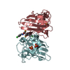
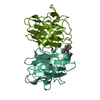
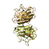
 Homo sapiens (human)
 X-RAY DIFFRACTION /
  Authors
 Citation
 Structure visualization
 Downloads & links
 Other downloads


 PDBj
PDBj


 Assembly
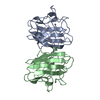


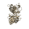
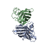
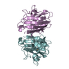
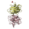
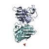
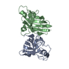
 Mass: 63.546 Da / Num. of mol.: 2 / Source method: obtained synthetically / Formula: Cu
 Mass: 65.409 Da / Num. of mol.: 2 / Source method: obtained synthetically / Formula: Zn
 Mass: 18.015 Da / Num. of mol.: 176 / Source method: isolated from a natural source / Formula: H2O
 Sample preparation
 Processing