 Movie
Movie Controller

[English] 日本語
 Yorodumi
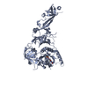
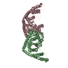
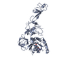
- PDB-1hyr: CRYSTAL STRUCTURE OF HUMAN MICA IN COMPLEX WITH NATURAL KILLER CE... -
+ Open data

- Basic information
| Entry | Database: PDB / ID: 1hyr | ||||||
|---|---|---|---|---|---|---|---|
| Title | CRYSTAL STRUCTURE OF HUMAN MICA IN COMPLEX WITH NATURAL KILLER CELL RECEPTOR NKG2D | ||||||
 Components Components |
| ||||||
 Keywords Keywords | IMMUNE SYSTEM / ACTIVATING NK CELL RECEPTOR / NKG2D / C-TYPE-LECTIN LIKE / MIC-A / MHC-I / COMPLEX | ||||||
| Function / homology |  Function and homology information Function and homology informationimmune response to tumor cell / negative regulation of natural killer cell chemotaxis / MHC class Ib receptor activity / negative regulation of natural killer cell activation / negative regulation of GTPase activity / natural killer cell lectin-like receptor binding / gamma-delta T cell activation / positive regulation of natural killer cell mediated cytotoxicity / negative regulation of natural killer cell mediated cytotoxicity / natural killer cell activation ...immune response to tumor cell / negative regulation of natural killer cell chemotaxis / MHC class Ib receptor activity / negative regulation of natural killer cell activation / negative regulation of GTPase activity / natural killer cell lectin-like receptor binding / gamma-delta T cell activation / positive regulation of natural killer cell mediated cytotoxicity / negative regulation of natural killer cell mediated cytotoxicity / natural killer cell activation / natural killer cell mediated cytotoxicity / stimulatory C-type lectin receptor signaling pathway / MHC class I protein binding / T cell costimulation / nitric oxide biosynthetic process / DAP12 interactions / T cell mediated cytotoxicity / Immunoregulatory interactions between a Lymphoid and a non-Lymphoid cell / positive regulation of nitric oxide biosynthetic process / positive regulation of type II interferon production / DAP12 signaling / cellular response to lipopolysaccharide / carbohydrate binding / response to heat / signaling receptor activity / killing of cells of another organism / defense response to virus / adaptive immune response / cell differentiation / defense response to bacterium / defense response to Gram-positive bacterium / immune response / receptor ligand activity / external side of plasma membrane / DNA damage response / cell surface / signal transduction / : / membrane / identical protein binding / plasma membrane / cytoplasm Similarity search - Function | ||||||
| Biological species |  Homo sapiens (human) Homo sapiens (human) | ||||||
| Method |  X-RAY DIFFRACTION / SYNCHROTRON / MAD / Resolution: 2.7 Å X-RAY DIFFRACTION / SYNCHROTRON / MAD / Resolution: 2.7 Å | ||||||
 Authors Authors | Li, P. / Strong, R.K. | ||||||
 Citation Citation | Journal: Nat.Immunol. / Year: 2001 Title: Complex structure of the activating immunoreceptor NKG2D and its MHC class I-like ligand MICA. Authors: Li, P. / Morris, D.L. / Willcox, B.E. / Steinle, A. / Spies, T. / Strong, R.K. #1: Journal: Immunity / Year: 1999Title: Crystal Structure of the MHC Class I Homolog Mic-A, a Gammadelta T Cell Ligand Authors: Li, P. / Willie, S.T. / Bauer, S. / Morris, D.L. / Spies, T. / Strong, R.K. #2: Journal: Science / Year: 1999Title: ACTIVATION OF NK CELLS AND T CELLS BY NKG2D, A RECEPTOR FOR STRESS INDUCIBLE MICA Authors: Bauer, S. / Groh, V. / Wu, J. / Steinle, A. / Phillips, J.H. / Lanier, L.L. / Spies, T. | ||||||
| History |
|
- Structure visualization
| Structure viewer | Molecule: MolmilJmol/JSmol |
|---|
- Downloads & links
-Download
| PDBx/mmCIF format | 1hyr.cif.gz | 109.9 KB | Display | PDBx/mmCIF format |
|---|---|---|---|---|
| PDB format | pdb1hyr.ent.gz | 85.7 KB | Display | PDB format |
| PDBx/mmJSON format | 1hyr.json.gz | Tree view | PDBx/mmJSON format | |
| Others |  Other downloads Other downloads |
-Validation report
| Arichive directory | https://data.pdbj.org/pub/pdb/validation_reports/hy/1hyrftp://data.pdbj.org/pub/pdb/validation_reports/hy/1hyr | HTTPS FTP |
|---|
-Related structure data
| Similar structure data |
|---|


-Links
 PDBj
PDBj


- Assembly
| Deposited unit | 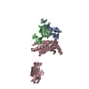
| ||||||||
|---|---|---|---|---|---|---|---|---|---|
| 1 |
| ||||||||
| Unit cell |
|
-Components
| #1: Protein | Mass: 15849.903 Da / Num. of mol.: 2 / Fragment: EXTRACELLULAR DOMAIN (RESIDUES 80 TO 216) Source method: isolated from a genetically manipulated source Source: (gene. exp.) Homo sapiens (human) / Cellular location: CELL SURFACE / Gene: NKG2D / Plasmid: PET22B / Species (production host): Escherichia coli / Production host:  #2: Protein | | Mass: 31731.434 Da / Num. of mol.: 1 / Fragment: EXTRACELLULAR DOMAIN (RESIDUES 1 TO 274) Source method: isolated from a genetically manipulated source Source: (gene. exp.) Homo sapiens (human) / Gene: MICA-001 / Plasmid: PET11 / Production host: #3: Water | ChemComp-HOH / |  Mass: 18.015 Da / Num. of mol.: 46 / Source method: isolated from a natural source / Formula: H2O Mass: 18.015 Da / Num. of mol.: 46 / Source method: isolated from a natural source / Formula: H2OHas protein modification | Y | |
|---|
-Experimental details
-Experiment
| Experiment | Method: X-RAY DIFFRACTION / Number of used crystals: 2 |
|---|
- Sample preparation
| Crystal | Density Matthews: 3.1 Å3/Da / Density % sol: 58.3 % | ||||||||||||||||||||||||||||||||||||||||||
|---|---|---|---|---|---|---|---|---|---|---|---|---|---|---|---|---|---|---|---|---|---|---|---|---|---|---|---|---|---|---|---|---|---|---|---|---|---|---|---|---|---|---|---|
| Crystal grow | Temperature: 295 K / Method: vapor diffusion, sitting drop / pH: 6.5 Details: (NH4)2SO4, pH 6.50, VAPOR DIFFUSION, SITTING DROP, temperature 295.0K | ||||||||||||||||||||||||||||||||||||||||||
| Crystal grow | *PLUS Temperature: 22 ℃ / pH: 7 | ||||||||||||||||||||||||||||||||||||||||||
| Components of the solutions | *PLUS
|
-Data collection
| Diffraction |
| ||||||||||||||||||
|---|---|---|---|---|---|---|---|---|---|---|---|---|---|---|---|---|---|---|---|
| Diffraction source |
| ||||||||||||||||||
| Detector | Type: ADSC QUANTUM 4 / Detector: CCD / Date: Oct 10, 2000 | ||||||||||||||||||
| Radiation |
| ||||||||||||||||||
| Radiation wavelength |
| ||||||||||||||||||
| Reflection | Resolution: 2.7→30 Å / Num. all: 256599 / Num. obs: 21938 / % possible obs: 99.9 % / Observed criterion σ(F): 0 / Observed criterion σ(I): 0 / Redundancy: 11.7 % / Biso Wilson estimate: 66.1 Å2 / Rmerge(I) obs: 0.076 / Rsym value: 0.076 / Net I/σ(I): 34 | ||||||||||||||||||
| Reflection shell | Resolution: 2.7→2.8 Å / Redundancy: 9 % / Rmerge(I) obs: 0.425 / Mean I/σ(I) obs: 4.1 / Num. unique all: 2150 / Rsym value: 0.425 / % possible all: 100 | ||||||||||||||||||
| Reflection | *PLUS Num. measured all: 256599 / Rmerge(I) obs: 0.076 | ||||||||||||||||||
| Reflection shell | *PLUS % possible obs: 100 % |

- Processing
| Software |
| |||||||||||||||||||||||||
|---|---|---|---|---|---|---|---|---|---|---|---|---|---|---|---|---|---|---|---|---|---|---|---|---|---|---|
| Refinement | Method to determine structure: MAD / Resolution: 2.7→29.78 Å / Rfactor Rfree error: 0.007 / Data cutoff high absF: 185946.74 / Data cutoff low absF: 0 / Isotropic thermal model: GROUP / Cross valid method: THROUGHOUT / σ(F): 0 / σ(I): 0 / Stereochemistry target values: Engh & Huber
| |||||||||||||||||||||||||
| Solvent computation | Solvent model: FLAT MODEL / Bsol: 33.93 Å2 / ksol: 0.302 e/Å3 | |||||||||||||||||||||||||
| Displacement parameters | Biso mean: 59.8 Å2
| |||||||||||||||||||||||||
| Refine analyze |
| |||||||||||||||||||||||||
| Refinement step | Cycle: LAST / Resolution: 2.7→29.78 Å
| |||||||||||||||||||||||||
| Refine LS restraints |
| |||||||||||||||||||||||||
| LS refinement shell | Resolution: 2.7→2.87 Å / Rfactor Rfree error: 0.025 / Total num. of bins used: 6
| |||||||||||||||||||||||||
| Xplor file |
| |||||||||||||||||||||||||
| Software | *PLUS Name: CNS / Version: 1 / Classification: refinement | |||||||||||||||||||||||||
| Refinement | *PLUS σ(F): 0 / % reflection Rfree: 7.4 % | |||||||||||||||||||||||||
| Solvent computation | *PLUS | |||||||||||||||||||||||||
| Displacement parameters | *PLUS Biso mean: 59.8 Å2 | |||||||||||||||||||||||||
| Refine LS restraints | *PLUS
| |||||||||||||||||||||||||
| LS refinement shell | *PLUS Rfactor Rfree: 0.397 / % reflection Rfree: 7.5 % / Rfactor Rwork: 0.346 |
