 Movie
Movie Controller

[English] 日本語
 Yorodumi
- PDB-1ml3: Evidences for a flip-flop catalytic mechanism of Trypanosoma cruz... -
+ Open data

- Basic information
| Entry | Database: PDB / ID: 1ml3 | ||||||
|---|---|---|---|---|---|---|---|
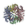
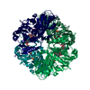
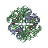
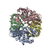
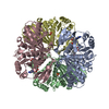
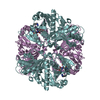
| Title | Evidences for a flip-flop catalytic mechanism of Trypanosoma cruzi glyceraldehyde-3-phosphate dehydrogenase, from its crystal structure in complex with reacted irreversible inhibitor 2-(2-phosphono-ethyl)-acrylic acid 4-nitro-phenyl ester | ||||||
 Components Components | Glyceraldehyde 3-phosphate dehydrogenase, glycosomal | ||||||
 Keywords Keywords | OXIDOREDUCTASE / protein covalent-inhibitor complex | ||||||
| Function / homology |  Function and homology information Function and homology informationglyceraldehyde-3-phosphate dehydrogenase (phosphorylating) / glyceraldehyde-3-phosphate dehydrogenase (NAD+) (phosphorylating) activity / glycosome / glycolytic process / glucose metabolic process / NAD binding / NADP binding / cytosol Similarity search - Function | ||||||
| Biological species |  | ||||||
| Method |  X-RAY DIFFRACTION / SYNCHROTRON / MOLECULAR REPLACEMENT / Resolution: 2.5 Å X-RAY DIFFRACTION / SYNCHROTRON / MOLECULAR REPLACEMENT / Resolution: 2.5 Å | ||||||
 Authors Authors | Castilho, M.S. / Pavao, F. / Oliva, G. | ||||||
 Citation Citation | Journal: Biochemistry / Year: 2003 Title: Evidence for the Two Phosphate Binding Sites of an Analogue of the Thioacyl Intermediate for the Trypanosoma cruzi Glyceraldehyde-3-phosphate Dehydrogenase-Catalyzed Reaction, from Its Crystal Structure. Authors: Castilho, M.S. / Pavao, F. / Oliva, G. / Ladame, S. / Willson, M. / Perie, J. | ||||||
| History |
| ||||||
| Remark 600 | HETEROGEN 2(2-phosphono-ethyl)-acrylic acid 4-nitro-phenyl ester underwent reaction with the ...HETEROGEN 2(2-phosphono-ethyl)-acrylic acid 4-nitro-phenyl ester underwent reaction with the protein. In chains B, C, and D, the product of the reaction, (3-FORMYL-BUT-3-ENYL)-PHOSPHONIC ACID, is bound to CYS 166. CYS 166 of chain A does not have this ligand bound. The ligand bound in alternate conformations for chains B and C. |
- Structure visualization
| Structure viewer | Molecule: MolmilJmol/JSmol |
|---|
- Downloads & links
-Download
| PDBx/mmCIF format | 1ml3.cif.gz | 298.9 KB | Display | PDBx/mmCIF format |
|---|---|---|---|---|
| PDB format | pdb1ml3.ent.gz | 240.8 KB | Display | PDB format |
| PDBx/mmJSON format | 1ml3.json.gz | Tree view | PDBx/mmJSON format | |
| Others |  Other downloads Other downloads |
-Validation report
| Summary document | 1ml3_validation.pdf.gz | 1.2 MB | Display | wwPDB validaton report |
|---|---|---|---|---|
| Full document | 1ml3_full_validation.pdf.gz | 1.2 MB | Display | |
| Data in XML | 1ml3_validation.xml.gz | 66.4 KB | Display | |
| Data in CIF | 1ml3_validation.cif.gz | 90 KB | Display | |
| Arichive directory | https://data.pdbj.org/pub/pdb/validation_reports/ml/1ml3ftp://data.pdbj.org/pub/pdb/validation_reports/ml/1ml3 | HTTPS FTP |
-Related structure data
| Related structure data | |
|---|---|
| Similar structure data |


-Links
 PDBj
PDBj
- Assembly
| Deposited unit | 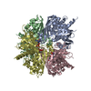
| ||||||||
|---|---|---|---|---|---|---|---|---|---|
| 1 |
| ||||||||
| Unit cell |
|
-Components
| #1: Protein | Mass: 39112.539 Da / Num. of mol.: 4 Source method: isolated from a genetically manipulated source Source: (gene. exp.)  References: UniProt: P22513, glyceraldehyde-3-phosphate dehydrogenase (phosphorylating) #2: Chemical |   Mass: 663.425 Da / Num. of mol.: 3 / Source method: obtained synthetically / Formula: C21H27N7O14P2 / Comment: NAD*YM Mass: 663.425 Da / Num. of mol.: 3 / Source method: obtained synthetically / Formula: C21H27N7O14P2 / Comment: NAD*YM#3: Chemical |   Mass: 164.096 Da / Num. of mol.: 3 / Source method: obtained synthetically / Formula: C5H9O4P Mass: 164.096 Da / Num. of mol.: 3 / Source method: obtained synthetically / Formula: C5H9O4P#4: Water | ChemComp-HOH / |  Mass: 18.015 Da / Num. of mol.: 695 / Source method: isolated from a natural source / Formula: H2O Mass: 18.015 Da / Num. of mol.: 695 / Source method: isolated from a natural source / Formula: H2OHas protein modification | Y | |
|---|
-Experimental details
-Experiment
| Experiment | Method: X-RAY DIFFRACTION / Number of used crystals: 1 |
|---|
- Sample preparation
| Crystal | Density Matthews: 2.32 Å3/Da / Density % sol: 47.03 % | ||||||||||||||||||||||||||||||||||||||||||
|---|---|---|---|---|---|---|---|---|---|---|---|---|---|---|---|---|---|---|---|---|---|---|---|---|---|---|---|---|---|---|---|---|---|---|---|---|---|---|---|---|---|---|---|
| Crystal grow | Temperature: 291 K / Method: vapor diffusion, hanging drop / pH: 7.5 Details: PEG 8000, CALCIUM ACETATE, EDTA, SODIUM AZIDE, pH 7.5, VAPOR DIFFUSION, HANGING DROP, temperature 291K | ||||||||||||||||||||||||||||||||||||||||||
| Crystal grow | *PLUS Temperature: 18 ℃ / Method: vapor diffusion, hanging drop / PH range low: 7.5 / PH range high: 7.3 | ||||||||||||||||||||||||||||||||||||||||||
| Components of the solutions | *PLUS
|
-Data collection
| Diffraction | Mean temperature: 100 K |
|---|---|
| Diffraction source | Source: SYNCHROTRON / Site: LNLS  / Beamline: D03B-MX1 / Wavelength: 1.5418 Å / Beamline: D03B-MX1 / Wavelength: 1.5418 Å |
| Detector | Type: MARRESEARCH / Detector: IMAGE PLATE / Date: Oct 5, 2000 |
| Radiation | Protocol: SINGLE WAVELENGTH / Monochromatic (M) / Laue (L): M / Scattering type: x-ray |
| Radiation wavelength | Wavelength: 1.5418 Å / Relative weight: 1 |
| Reflection | Resolution: 2.5→20 Å / Num. obs: 48450 / % possible obs: 97.5 % / Redundancy: 2.77 % / Rmerge(I) obs: 0.113 / Net I/σ(I): 9.73 |
| Reflection shell | Resolution: 2.5→2.56 Å / Redundancy: 2.47 % / Rmerge(I) obs: 0.438 / Mean I/σ(I) obs: 2.57 / % possible all: 99 |
| Reflection | *PLUS % possible obs: 99 % / Redundancy: 2.8 % / Num. measured all: 134297 |
| Reflection shell | *PLUS % possible obs: 97.5 % / Redundancy: 2.5 % / Mean I/σ(I) obs: 2.6 |
- Processing
| Software |
| ||||||||||||||||||||
|---|---|---|---|---|---|---|---|---|---|---|---|---|---|---|---|---|---|---|---|---|---|
| Refinement | Method to determine structure: MOLECULAR REPLACEMENT Starting model: NATIVE STRUCTURE OF T.cruzi gGAPDH (Souza et al FEBES letters (1998), 424, 131-135) Resolution: 2.5→20 Å
| ||||||||||||||||||||
| Refinement step | Cycle: LAST / Resolution: 2.5→20 Å
| ||||||||||||||||||||
| Refine LS restraints |
| ||||||||||||||||||||
| Refinement | *PLUS % reflection Rfree: 3 % / Rfactor Rfree: 0.26 / Rfactor Rwork: 0.2 | ||||||||||||||||||||
| Solvent computation | *PLUS | ||||||||||||||||||||
| Displacement parameters | *PLUS | ||||||||||||||||||||
| Refine LS restraints | *PLUS
|
