 Movie
Movie Controller

[English] 日本語
 Yorodumi
- PDB-1gyp: CRYSTAL STRUCTURE OF GLYCOSOMAL GLYCERALDEHYDE-3-PHOSPHATE DEHYDR... -
+ Open data

- Basic information
| Entry | Database: PDB / ID: 1gyp | ||||||
|---|---|---|---|---|---|---|---|
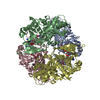
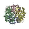
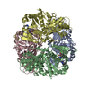
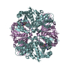
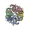
| Title | CRYSTAL STRUCTURE OF GLYCOSOMAL GLYCERALDEHYDE-3-PHOSPHATE DEHYDROGENASE FROM LEISHMANIA MEXICANA: IMPLICATIONS FOR STRUCTURE-BASED DRUG DESIGN AND A NEW POSITION FOR THE INORGANIC PHOSPHATE BINDING SITE | ||||||
 Components Components | GLYCERALDEHYDE-3-PHOSPHATE DEHYDROGENASE | ||||||
 Keywords Keywords | OXIDOREDUCTASE (ALDEHYDE(D)-NAD+(A)) | ||||||
| Function / homology |  Function and homology information Function and homology informationglyceraldehyde-3-phosphate dehydrogenase (phosphorylating) / glycosome / glyceraldehyde-3-phosphate dehydrogenase (NAD+) (phosphorylating) activity / glycolytic process / glucose metabolic process / NAD binding / NADP binding / cytosol Similarity search - Function | ||||||
| Biological species |   Leishmania mexicana (eukaryote) Leishmania mexicana (eukaryote) | ||||||
| Method |  X-RAY DIFFRACTION / SYNCHROTRON / Resolution: 2.8 Å X-RAY DIFFRACTION / SYNCHROTRON / Resolution: 2.8 Å | ||||||
 Authors Authors | Kim, H. / Feil, I.K. / Verlinde, C.L.M.J. / Petra, P.H. / Hol, W.G.J. | ||||||
 Citation Citation | Journal: Biochemistry / Year: 1995 Title: Crystal structure of glycosomal glyceraldehyde-3-phosphate dehydrogenase from Leishmania mexicana: implications for structure-based drug design and a new position for the inorganic phosphate binding site. Authors: Kim, H. / Feil, I.K. / Verlinde, C.L. / Petra, P.H. / Hol, W.G. | ||||||
| History |
|
- Structure visualization
| Structure viewer | Molecule: MolmilJmol/JSmol |
|---|
- Downloads & links
-Download
| PDBx/mmCIF format | 1gyp.cif.gz | 275.4 KB | Display | PDBx/mmCIF format |
|---|---|---|---|---|
| PDB format | pdb1gyp.ent.gz | 228.1 KB | Display | PDB format |
| PDBx/mmJSON format | 1gyp.json.gz | Tree view | PDBx/mmJSON format | |
| Others |  Other downloads Other downloads |
-Validation report
| Arichive directory | https://data.pdbj.org/pub/pdb/validation_reports/gy/1gypftp://data.pdbj.org/pub/pdb/validation_reports/gy/1gyp | HTTPS FTP |
|---|
-Related structure data
| Similar structure data |
|---|


-Links
 PDBj
PDBj
- Assembly
| Deposited unit | 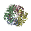
| ||||||||||||||||
|---|---|---|---|---|---|---|---|---|---|---|---|---|---|---|---|---|---|
| 1 |
| ||||||||||||||||
| Unit cell |
| ||||||||||||||||
| Noncrystallographic symmetry (NCS) | NCS oper:
| ||||||||||||||||
| Details | THERE IS ONE 222 HOMOTETRAMER PER ASYMMETRIC UNIT RESULTING IN NON-CRYSTALLOGRAPHIC 222 SYMMETRY WITHIN THE ASYMMETRIC UNIT. |
-Components
| #1: Protein | Mass: 38693.129 Da / Num. of mol.: 4 Source method: isolated from a genetically manipulated source Source: (gene. exp.) Leishmania mexicana (eukaryote) / Gene: GLYCERALDEHYDE-3-PHOSPHATE / Plasmid: PET-3AGene (production host): GLYCERALDEHYDE-3-PHOSPHATE DEHYDROGENASE Production host:  References: UniProt: Q27890, glyceraldehyde-3-phosphate dehydrogenase (phosphorylating) #2: Chemical | ChemComp-PO4 /   Mass: 94.971 Da / Num. of mol.: 8 / Source method: obtained synthetically / Formula: PO4 Mass: 94.971 Da / Num. of mol.: 8 / Source method: obtained synthetically / Formula: PO4#3: Chemical | ChemComp-NAD /   Mass: 663.425 Da / Num. of mol.: 4 / Source method: obtained synthetically / Formula: C21H27N7O14P2 / Comment: NAD*YM Mass: 663.425 Da / Num. of mol.: 4 / Source method: obtained synthetically / Formula: C21H27N7O14P2 / Comment: NAD*YM |
|---|
-Experimental details
-Experiment
| Experiment | Method: X-RAY DIFFRACTION |
|---|
- Sample preparation
| Crystal | Density Matthews: 2.81 Å3/Da / Density % sol: 56.21 % | ||||||||||||||||||||
|---|---|---|---|---|---|---|---|---|---|---|---|---|---|---|---|---|---|---|---|---|---|
| Crystal grow | *PLUS pH: 7 / Method: vapor diffusion, sitting drop / Details: pH is adjusted with 0.2 M citric acid | ||||||||||||||||||||
| Components of the solutions | *PLUS
|
-Data collection
| Diffraction source | Source: SYNCHROTRON / Site: SSRL  / Beamline: BL7-1 / Wavelength: 1.08 / Beamline: BL7-1 / Wavelength: 1.08 |
|---|---|
| Detector | Type: MARRESEARCH / Detector: IMAGE PLATE / Date: Mar 25, 1994 |
| Radiation | Monochromatic (M) / Laue (L): M / Scattering type: x-ray |
| Radiation wavelength | Wavelength: 1.08 Å / Relative weight: 1 |
| Reflection | Num. obs: 37522 / % possible obs: 86 % / Observed criterion σ(I): 2 / Redundancy: 3 % / Rmerge(I) obs: 0.076 |
| Reflection | *PLUS Highest resolution: 2.8 Å / Lowest resolution: 30 Å / Rmerge(I) obs: 0.076 |
| Reflection shell | *PLUS Highest resolution: 2.8 Å / Lowest resolution: 2.87 Å / % possible obs: 77 % / Rmerge(I) obs: 0.128 |
- Processing
| Software |
| ||||||||||||||||||||||||||||||||||||||||||||||||||||||||||||
|---|---|---|---|---|---|---|---|---|---|---|---|---|---|---|---|---|---|---|---|---|---|---|---|---|---|---|---|---|---|---|---|---|---|---|---|---|---|---|---|---|---|---|---|---|---|---|---|---|---|---|---|---|---|---|---|---|---|---|---|---|---|
| Refinement | Resolution: 2.8→10 Å / σ(F): 2 /
| ||||||||||||||||||||||||||||||||||||||||||||||||||||||||||||
| Displacement parameters | Biso mean: 33 Å2 | ||||||||||||||||||||||||||||||||||||||||||||||||||||||||||||
| Refinement step | Cycle: LAST / Resolution: 2.8→10 Å
| ||||||||||||||||||||||||||||||||||||||||||||||||||||||||||||
| Refine LS restraints |
|
