 Movie
Movie Controller

[English] 日本語
 Yorodumi
- PDB-2x5j: Crystal structure of the Apoform of the D-Erythrose-4-phosphate d... -
+ Open data

- Basic information
| Entry | Database: PDB / ID: 2x5j | ||||||
|---|---|---|---|---|---|---|---|
| Title | Crystal structure of the Apoform of the D-Erythrose-4-phosphate dehydrogenase from E. coli | ||||||
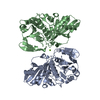
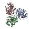
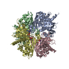
 Components Components | D-ERYTHROSE-4-PHOSPHATE DEHYDROGENASE | ||||||
 Keywords Keywords | OXIDOREDUCTASE / HYDRIDE TRANSFER / ALDEHYDE DEHYDROGENASE / PYRIDOXINE BIOSYNTHESIS | ||||||
| Function / homology |  Function and homology information Function and homology informationerythrose-4-phosphate dehydrogenase / erythrose-4-phosphate dehydrogenase activity / pyridoxal 5'-phosphate biosynthetic process / pyridoxine biosynthetic process / glyceraldehyde-3-phosphate dehydrogenase (NAD+) (phosphorylating) activity / glucose metabolic process / NAD binding / cytosol Similarity search - Function | ||||||
| Biological species |  | ||||||
| Method |  X-RAY DIFFRACTION / SYNCHROTRON / MOLECULAR REPLACEMENT / Resolution: 2.3 Å X-RAY DIFFRACTION / SYNCHROTRON / MOLECULAR REPLACEMENT / Resolution: 2.3 Å | ||||||
 Authors Authors | Moniot, S. / Didierjean, C. / Boschi-Muller, S. / Branlant, G. / Corbier, C. | ||||||
 Citation Citation | Journal: To be Published Title: Structural Characterization of Erythrose-4- Phosphate Dehydrogenase from Escherichia Coli: Peculiar Features When Compared to Phosphorylating Gapdhs Authors: Moniot, S. / Didierjean, C. / Boschi-Muller, S. / Branlant, G. / Corbier, C. | ||||||
| History |
|
- Structure visualization
| Structure viewer | Molecule: MolmilJmol/JSmol |
|---|
- Downloads & links
-Download
| PDBx/mmCIF format | 2x5j.cif.gz | 268 KB | Display | PDBx/mmCIF format |
|---|---|---|---|---|
| PDB format | pdb2x5j.ent.gz | 219.2 KB | Display | PDB format |
| PDBx/mmJSON format | 2x5j.json.gz | Tree view | PDBx/mmJSON format | |
| Others |  Other downloads Other downloads |
-Validation report
| Arichive directory | https://data.pdbj.org/pub/pdb/validation_reports/x5/2x5jftp://data.pdbj.org/pub/pdb/validation_reports/x5/2x5j | HTTPS FTP |
|---|
-Related structure data
| Related structure data |  2x5kC  2xf8C 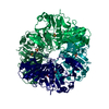 2gd1S  2x5m C: citing same article ( S: Starting model for refinement |
|---|---|
| Similar structure data |


-Links
 PDBj
PDBj
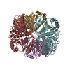
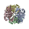
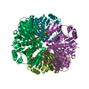
- Assembly
| Deposited unit | 
| ||||||||||||||||
|---|---|---|---|---|---|---|---|---|---|---|---|---|---|---|---|---|---|
| 1 |
| ||||||||||||||||
| Unit cell |
| ||||||||||||||||
| Noncrystallographic symmetry (NCS) | NCS oper:
|
-Components
| #1: Protein | Mass: 37347.328 Da / Num. of mol.: 4 Source method: isolated from a genetically manipulated source Details: COMPLEX WITH PHOSPHATE ANION / Source: (gene. exp.) References: UniProt: P0A9B6, erythrose-4-phosphate dehydrogenase #2: Chemical | ChemComp-PO4 /   Mass: 94.971 Da / Num. of mol.: 4 / Source method: obtained synthetically / Formula: PO4 Mass: 94.971 Da / Num. of mol.: 4 / Source method: obtained synthetically / Formula: PO4#3: Water | ChemComp-HOH / |  Mass: 18.015 Da / Num. of mol.: 539 / Source method: isolated from a natural source / Formula: H2O Mass: 18.015 Da / Num. of mol.: 539 / Source method: isolated from a natural source / Formula: H2OHas protein modification | Y | |
|---|
-Experimental details
-Experiment
| Experiment | Method: X-RAY DIFFRACTION / Number of used crystals: 1 |
|---|
- Sample preparation
| Crystal | Density Matthews: 2.18 Å3/Da / Density % sol: 43.13 % / Description: NONE |
|---|---|
| Crystal grow | Details: 10 % (W/V) PEG 8000, 100 MM NA/K PHOSPHATE BUFFER PH 6.2, 200 MM NACL |
-Data collection
| Diffraction | Mean temperature: 100 K |
|---|---|
| Diffraction source | Source: SYNCHROTRON / Site: ESRF  / Beamline: BM30A / Wavelength: 0.9797 / Beamline: BM30A / Wavelength: 0.9797 |
| Detector | Type: MARRESEARCH / Detector: CCD |
| Radiation | Protocol: SINGLE WAVELENGTH / Monochromatic (M) / Laue (L): M / Scattering type: x-ray |
| Radiation wavelength | Wavelength: 0.9797 Å / Relative weight: 1 |
| Reflection | Resolution: 2.3→34.5 Å / Num. obs: 59159 / % possible obs: 99.6 % / Observed criterion σ(I): -3 / Redundancy: 5.6 % / Rmerge(I) obs: 0.06 / Net I/σ(I): 24.4 |
| Reflection shell | Resolution: 2.3→2.42 Å / Redundancy: 5.5 % / Rmerge(I) obs: 0.43 / Mean I/σ(I) obs: 5.6 / % possible all: 99.8 |
- Processing
| Software |
| ||||||||||||||||||||||||||||||||||||||||||||||||||||||||||||||||||||||||||||||||||||||||||||||||||||||||||||||||||||||||||||||||||||||||||||||||||||||||||||||||||||||||||||||||||||||
|---|---|---|---|---|---|---|---|---|---|---|---|---|---|---|---|---|---|---|---|---|---|---|---|---|---|---|---|---|---|---|---|---|---|---|---|---|---|---|---|---|---|---|---|---|---|---|---|---|---|---|---|---|---|---|---|---|---|---|---|---|---|---|---|---|---|---|---|---|---|---|---|---|---|---|---|---|---|---|---|---|---|---|---|---|---|---|---|---|---|---|---|---|---|---|---|---|---|---|---|---|---|---|---|---|---|---|---|---|---|---|---|---|---|---|---|---|---|---|---|---|---|---|---|---|---|---|---|---|---|---|---|---|---|---|---|---|---|---|---|---|---|---|---|---|---|---|---|---|---|---|---|---|---|---|---|---|---|---|---|---|---|---|---|---|---|---|---|---|---|---|---|---|---|---|---|---|---|---|---|---|---|---|---|
| Refinement | Method to determine structure: MOLECULAR REPLACEMENT Starting model: PDB ENTRY 2GD1 Resolution: 2.3→34.5 Å / Cor.coef. Fo:Fc: 0.938 / Cor.coef. Fo:Fc free: 0.899 / SU B: 8.315 / SU ML: 0.202 / Cross valid method: THROUGHOUT / ESU R: 0.441 / ESU R Free: 0.273 / Stereochemistry target values: MAXIMUM LIKELIHOOD Details: HYDROGENS HAVE BEEN ADDED IN THE RIDING POSITIONS. U VALUES REFINED INDIVIDUALLY
| ||||||||||||||||||||||||||||||||||||||||||||||||||||||||||||||||||||||||||||||||||||||||||||||||||||||||||||||||||||||||||||||||||||||||||||||||||||||||||||||||||||||||||||||||||||||
| Solvent computation | Ion probe radii: 0.8 Å / Shrinkage radii: 0.8 Å / VDW probe radii: 1.4 Å / Solvent model: MASK | ||||||||||||||||||||||||||||||||||||||||||||||||||||||||||||||||||||||||||||||||||||||||||||||||||||||||||||||||||||||||||||||||||||||||||||||||||||||||||||||||||||||||||||||||||||||
| Displacement parameters | Biso mean: 33.711 Å2
| ||||||||||||||||||||||||||||||||||||||||||||||||||||||||||||||||||||||||||||||||||||||||||||||||||||||||||||||||||||||||||||||||||||||||||||||||||||||||||||||||||||||||||||||||||||||
| Refinement step | Cycle: LAST / Resolution: 2.3→34.5 Å
| ||||||||||||||||||||||||||||||||||||||||||||||||||||||||||||||||||||||||||||||||||||||||||||||||||||||||||||||||||||||||||||||||||||||||||||||||||||||||||||||||||||||||||||||||||||||
| Refine LS restraints |
|