 Movie
Movie Controller

[English] 日本語
 Yorodumi
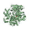
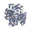
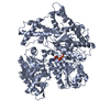
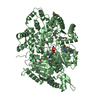
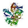
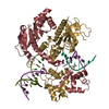
- PDB-1mkf: VIRAL CHEMOKINE BINDING PROTEIN M3 FROM MURINE GAMMAHERPESVIRUS 68 -
+ Open data

- Basic information
| Entry | Database: PDB / ID: 1mkf | ||||||
|---|---|---|---|---|---|---|---|
| Title | VIRAL CHEMOKINE BINDING PROTEIN M3 FROM MURINE GAMMAHERPESVIRUS 68 | ||||||
 Components Components | M3 | ||||||
 Keywords Keywords | IMMUNE SYSTEM / HERPESVIRUS / VIRAL IMMUNE EVASION / CHEMOKINE BINDING PROTEIN / DECOY RECEPTOR / Structural Genomics / PSI / Protein Structure Initiative / Midwest Center for Structural Genomics / MCSG | ||||||
| Function / homology |  Function and homology information Function and homology information | ||||||
| Biological species |  Murid herpesvirus 4 (Murine herpesvirus 68) Murid herpesvirus 4 (Murine herpesvirus 68) | ||||||
| Method |  X-RAY DIFFRACTION / SYNCHROTRON / MIR / Resolution: 2.1 Å X-RAY DIFFRACTION / SYNCHROTRON / MIR / Resolution: 2.1 Å | ||||||
 Authors Authors | Alexander, J.M. / Fremont, D.H. / Midwest Center for Structural Genomics (MCSG) | ||||||
 Citation Citation | Journal: Cell(Cambridge,Mass.) / Year: 2002 Title: Structural Basis of Chemokine Sequestration by a Herpesvirus Decoy Receptor Authors: Alexander, J.M. / Nelson, C.A. / Van Berkel, V. / Lau, E.K. / Studts, J.M. / Brett, T.J. / Speck, S.H. / Handel, T.M. / Virgin, H.W. / Fremont, D.H. | ||||||
| History |
|
- Structure visualization
| Structure viewer | Molecule: MolmilJmol/JSmol |
|---|
- Downloads & links
-Download
| PDBx/mmCIF format | 1mkf.cif.gz | 158.6 KB | Display | PDBx/mmCIF format |
|---|---|---|---|---|
| PDB format | pdb1mkf.ent.gz | 126.2 KB | Display | PDB format |
| PDBx/mmJSON format | 1mkf.json.gz | Tree view | PDBx/mmJSON format | |
| Others |  Other downloads Other downloads |
-Validation report
| Summary document | 1mkf_validation.pdf.gz | 372.7 KB | Display | wwPDB validaton report |
|---|---|---|---|---|
| Full document | 1mkf_full_validation.pdf.gz | 386.5 KB | Display | |
| Data in XML | 1mkf_validation.xml.gz | 16.6 KB | Display | |
| Data in CIF | 1mkf_validation.cif.gz | 26.7 KB | Display | |
| Arichive directory | https://data.pdbj.org/pub/pdb/validation_reports/mk/1mkfftp://data.pdbj.org/pub/pdb/validation_reports/mk/1mkf | HTTPS FTP |
-Related structure data


-Links
 PDBj
PDBj- Assembly
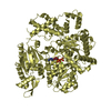
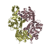
| Deposited unit | 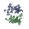
| ||||||||
|---|---|---|---|---|---|---|---|---|---|
| 1 |
| ||||||||
| 2 | 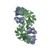
| ||||||||
| Unit cell |
|
-Components
| #1: Protein | Mass: 41826.230 Da / Num. of mol.: 2 Source method: isolated from a genetically manipulated source Source: (gene. exp.) Murid herpesvirus 4 (Murine herpesvirus 68)Genus: Rhadinovirus / Gene: M3 / Plasmid: pFB-1 / Cell line (production host): SF9 / Production host:   Spodoptera frugiperda (fall armyworm) / References: UniProt: O41925 Spodoptera frugiperda (fall armyworm) / References: UniProt: O41925#2: Water | ChemComp-HOH / |  Mass: 18.015 Da / Num. of mol.: 332 / Source method: isolated from a natural source / Formula: H2O Mass: 18.015 Da / Num. of mol.: 332 / Source method: isolated from a natural source / Formula: H2OHas protein modification | Y | |
|---|
-Experimental details
-Experiment
| Experiment | Method: X-RAY DIFFRACTION / Number of used crystals: 1 |
|---|
- Sample preparation
| Crystal | Density Matthews: 2.66 Å3/Da / Density % sol: 53 % | |||||||||||||||||||||||||||||||||||
|---|---|---|---|---|---|---|---|---|---|---|---|---|---|---|---|---|---|---|---|---|---|---|---|---|---|---|---|---|---|---|---|---|---|---|---|---|
| Crystal grow | Temperature: 293 K / Method: vapor diffusion, hanging drop / pH: 5.1 Details: 18% PEG4000, 100MM CACL2, 100MM IMIDAZOLE/MALIC ACID PH 5.1, pH 5.10, VAPOR DIFFUSION, HANGING DROP, temperature 293K | |||||||||||||||||||||||||||||||||||
| Crystal grow | *PLUS pH: 5.1 | |||||||||||||||||||||||||||||||||||
| Components of the solutions | *PLUS
|
-Data collection
| Diffraction | Mean temperature: 110 K |
|---|---|
| Diffraction source | Source: SYNCHROTRON / Site: NSLS  / Beamline: X25 / Wavelength: 1.1 / Wavelength: 1.1 Å / Beamline: X25 / Wavelength: 1.1 / Wavelength: 1.1 Å |
| Detector | Type: ADSC QUANTUM 4 / Detector: CCD / Date: Jun 1, 2000 |
| Radiation | Monochromator: SI 111 CHANNEL / Protocol: SINGLE WAVELENGTH / Monochromatic (M) / Laue (L): M / Scattering type: x-ray |
| Radiation wavelength | Wavelength: 1.1 Å / Relative weight: 1 |
| Reflection | Resolution: 2.1→20 Å / Num. all: 43484 / Num. obs: 43484 / % possible obs: 98 % / Observed criterion σ(F): 0 / Observed criterion σ(I): -3 / Redundancy: 3.1 % / Biso Wilson estimate: 40.6 Å2 / Rsym value: 0.052 / Net I/σ(I): 20.4 |
| Reflection shell | Resolution: 2.1→2.2 Å / Mean I/σ(I) obs: 2.9 / Rsym value: 0.346 / % possible all: 97 |
| Reflection | *PLUS Highest resolution: 2.1 Å / Lowest resolution: 20 Å / Redundancy: 3.1 % / Num. measured all: 133215 / Rmerge(I) obs: 0.052 |
| Reflection shell | *PLUS Highest resolution: 2.1 Å / Lowest resolution: 2.2 Å / % possible obs: 97 % / Rmerge(I) obs: 0.346 |
- Processing
| Software |
| ||||||||||||||||||||||||||||||||||||
|---|---|---|---|---|---|---|---|---|---|---|---|---|---|---|---|---|---|---|---|---|---|---|---|---|---|---|---|---|---|---|---|---|---|---|---|---|---|
| Refinement | Method to determine structure: MIR / Resolution: 2.1→20 Å / Rfactor Rfree error: 0.006 / Data cutoff high absF: 25464.2 / Data cutoff high rms absF: 25464.2 / Data cutoff low absF: 0 / Isotropic thermal model: RESTRAINED / Cross valid method: THROUGHOUT / σ(F): 0 / Stereochemistry target values: Engh & Huber
| ||||||||||||||||||||||||||||||||||||
| Solvent computation | Bsol: 57.7995 Å2 / ksol: 0.341932 e/Å3 | ||||||||||||||||||||||||||||||||||||
| Displacement parameters | Biso mean: 47.8 Å2
| ||||||||||||||||||||||||||||||||||||
| Refine analyze |
| ||||||||||||||||||||||||||||||||||||
| Refinement step | Cycle: LAST / Resolution: 2.1→20 Å
| ||||||||||||||||||||||||||||||||||||
| Refine LS restraints |
| ||||||||||||||||||||||||||||||||||||
| LS refinement shell | Resolution: 2.1→2.2 Å / Rfactor Rfree error: 0.024 / Total num. of bins used: 8
| ||||||||||||||||||||||||||||||||||||
| Xplor file |
| ||||||||||||||||||||||||||||||||||||
| Refinement | *PLUS Highest resolution: 2.1 Å / Lowest resolution: 20 Å / Num. reflection Rfree: 2506 | ||||||||||||||||||||||||||||||||||||
| Solvent computation | *PLUS | ||||||||||||||||||||||||||||||||||||
| Displacement parameters | *PLUS | ||||||||||||||||||||||||||||||||||||
| Refine LS restraints | *PLUS
| ||||||||||||||||||||||||||||||||||||
| LS refinement shell | *PLUS Highest resolution: 2.1 Å / Lowest resolution: 2.2 Å |
