 Movie
Movie Controller


+ Open data

- Basic information
| Entry | Database: PDB / ID: 1dok | ||||||
|---|---|---|---|---|---|---|---|
| Title | MONOCYTE CHEMOATTRACTANT PROTEIN 1, P-FORM | ||||||
 Components Components | MONOCYTE CHEMOATTRACTANT PROTEIN 1 | ||||||
 Keywords Keywords | CHEMOATTRACTANT / CYTOKINE | ||||||
| Function / homology |  Function and homology information Function and homology informationhelper T cell extravasation / chemokine (C-C motif) ligand 2 signaling pathway / CCR2 chemokine receptor binding / negative regulation of natural killer cell chemotaxis / astrocyte cell migration / chemokine receptor binding / ATF4 activates genes in response to endoplasmic reticulum stress / CCR chemokine receptor binding / negative regulation of glial cell apoptotic process / positive regulation of apoptotic cell clearance ...helper T cell extravasation / chemokine (C-C motif) ligand 2 signaling pathway / CCR2 chemokine receptor binding / negative regulation of natural killer cell chemotaxis / astrocyte cell migration / chemokine receptor binding / ATF4 activates genes in response to endoplasmic reticulum stress / CCR chemokine receptor binding / negative regulation of glial cell apoptotic process / positive regulation of apoptotic cell clearance / NFE2L2 regulating inflammation associated genes / eosinophil chemotaxis / cellular homeostasis / positive regulation of glutamate receptor signaling pathway / negative regulation of vascular endothelial cell proliferation / chemokine activity / Chemokine receptors bind chemokines / negative regulation of G1/S transition of mitotic cell cycle / positive regulation of calcium ion import / chemoattractant activity / positive regulation of macrophage chemotaxis / Interleukin-10 signaling / macrophage chemotaxis / humoral immune response / monocyte chemotaxis / positive regulation of endothelial cell apoptotic process / cellular response to interleukin-1 / G protein-coupled receptor signaling pathway, coupled to cyclic nucleotide second messenger / cell surface receptor signaling pathway via JAK-STAT / positive regulation of synaptic transmission, glutamatergic / cellular response to fibroblast growth factor stimulus / cytoskeleton organization / sensory perception of pain / viral genome replication / animal organ morphogenesis / chemokine-mediated signaling pathway / response to bacterium / cellular response to type II interferon / cellular response to tumor necrosis factor / positive regulation of T cell activation / chemotaxis / cytokine-mediated signaling pathway / regulation of cell shape / antimicrobial humoral immune response mediated by antimicrobial peptide / cellular response to lipopolysaccharide / positive regulation of cytosolic calcium ion concentration / Interleukin-4 and Interleukin-13 signaling / angiogenesis / protein phosphorylation / negative regulation of neuron apoptotic process / protein kinase activity / cell surface receptor signaling pathway / cell adhesion / positive regulation of cell migration / G protein-coupled receptor signaling pathway / inflammatory response / signaling receptor binding / positive regulation of gene expression / signal transduction / : / extracellular region / membrane Similarity search - Function | ||||||
| Biological species |  Homo sapiens (human) Homo sapiens (human) | ||||||
| Method |  X-RAY DIFFRACTION / MOLECULAR REPLACEMENT / Resolution: 1.85 Å X-RAY DIFFRACTION / MOLECULAR REPLACEMENT / Resolution: 1.85 Å | ||||||
 Authors Authors | Lubkowski, J. / Bujacz, G. / Boque, L. / Wlodawer, A. / Domaille, P.J. / Handel, T.M. | ||||||
 Citation Citation | Journal: Nat.Struct.Biol. / Year: 1997 Title: The structure of MCP-1 in two crystal forms provides a rare example of variable quaternary interactions. Authors: Lubkowski, J. / Bujacz, G. / Boque, L. / Domaille, P.J. / Handel, T.M. / Wlodawer, A. #1: Journal: Biochemistry / Year: 1996Title: Heteronuclear (1H, 13C, 15N) NMR Assignments and Solution Structure of the Monocyte Chemoattractant Protein-1 (Mcp-1) Dimer Authors: Handel, T.M. / Domaille, P.J. | ||||||
| History |
|
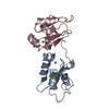
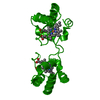
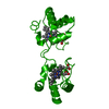
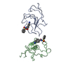
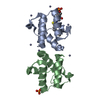
- Structure visualization
| Structure viewer | Molecule: MolmilJmol/JSmol |
|---|
- Downloads & links
-Download
| PDBx/mmCIF format | 1dok.cif.gz | 44.8 KB | Display | PDBx/mmCIF format |
|---|---|---|---|---|
| PDB format | pdb1dok.ent.gz | 31.8 KB | Display | PDB format |
| PDBx/mmJSON format | 1dok.json.gz | Tree view | PDBx/mmJSON format | |
| Others |  Other downloads Other downloads |
-Validation report
| Arichive directory | https://data.pdbj.org/pub/pdb/validation_reports/do/1dokftp://data.pdbj.org/pub/pdb/validation_reports/do/1dok | HTTPS FTP |
|---|
-Related structure data
| Related structure data |  1dolC  1domS S: Starting model for refinement C: citing same article ( |
|---|---|
| Similar structure data |


-Links
 PDBj
PDBj


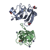
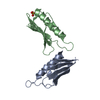
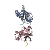
- Assembly
| Deposited unit | 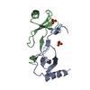
| ||||||||
|---|---|---|---|---|---|---|---|---|---|
| 1 |
| ||||||||
| 2 | 
| ||||||||
| Unit cell |
| ||||||||
| Noncrystallographic symmetry (NCS) | NCS oper: (Code: given Matrix: (-0.652315, -0.596164, 0.468052), Vector: |
-Components
| #1: Protein | Mass: 8830.239 Da / Num. of mol.: 2 Mutation: N-TERMINAL METHIONINE NOT REMOVED POST-TRANSLATIONALLY Source method: isolated from a genetically manipulated source Source: (gene. exp.) Homo sapiens (human) / Plasmid: BL21 / Production host:  #2: Chemical |   Mass: 96.063 Da / Num. of mol.: 2 / Source method: obtained synthetically / Formula: SO4 Mass: 96.063 Da / Num. of mol.: 2 / Source method: obtained synthetically / Formula: SO4#3: Water | ChemComp-HOH / |  Mass: 18.015 Da / Num. of mol.: 116 / Source method: isolated from a natural source / Formula: H2O Mass: 18.015 Da / Num. of mol.: 116 / Source method: isolated from a natural source / Formula: H2OHas protein modification | Y | Sequence details | RESIDUE MET 0 IS AN ARTIFACT OF EXPRESSION | |
|---|
-Experimental details
-Experiment
| Experiment | Method: X-RAY DIFFRACTION / Number of used crystals: 1 |
|---|
- Sample preparation
| Crystal | Density Matthews: 2.04 Å3/Da / Density % sol: 32 % Description: INITIAL MODEL FOR MOLECULAR REPLACEMENT WAS MODIFIED AS DESCRIBED IN JOURNAL ARTICLE. | ||||||||||||||||||||
|---|---|---|---|---|---|---|---|---|---|---|---|---|---|---|---|---|---|---|---|---|---|
| Crystal grow | Method: vapor diffusion, hanging drop / pH: 8 Details: 10 MG/ML PROTEIN IN 50 MM TRIS BUFFER PH 7.5-8 EQUILIBRATED AGAINST 50-55% AMMONIUM SULFATE USING HANGING DROP VAPOR DIFFUSION METHOD., pH 8.0, vapor diffusion - hanging drop PH range: 7.5-8.0 | ||||||||||||||||||||
| Crystal grow | *PLUS Method: vapor diffusion | ||||||||||||||||||||
| Components of the solutions | *PLUS
|
-Data collection
| Diffraction | Mean temperature: 298 K |
|---|---|
| Diffraction source | Source: ROTATING ANODE / Type: RIGAKU RUH2R / Wavelength: 1.5418 |
| Detector | Type: RIGAKU / Detector: IMAGE PLATE / Date: Nov 5, 1995 / Details: COLLIMATOR |
| Radiation | Monochromator: GRAPHITE(002) / Monochromatic (M) / Laue (L): M / Scattering type: x-ray |
| Radiation wavelength | Wavelength: 1.5418 Å / Relative weight: 1 |
| Reflection | Resolution: 1.85→10 Å / Num. obs: 12318 / % possible obs: 97 % / Observed criterion σ(I): 0 / Redundancy: 4.59 % / Rsym value: 0.078 / Net I/σ(I): 15.3 |
| Reflection shell | Resolution: 1.85→1.92 Å / Redundancy: 3.82 % / Mean I/σ(I) obs: 2.15 / Rsym value: 0.363 / % possible all: 94.7 |
| Reflection | *PLUS Rmerge(I) obs: 0.078 |
| Reflection shell | *PLUS % possible obs: 94.7 % / Rmerge(I) obs: 0.363 |
- Processing
| Software |
| ||||||||||||||||||||||||||||||||||||||||||||||||||||||||||||||||||||||||||||||||
|---|---|---|---|---|---|---|---|---|---|---|---|---|---|---|---|---|---|---|---|---|---|---|---|---|---|---|---|---|---|---|---|---|---|---|---|---|---|---|---|---|---|---|---|---|---|---|---|---|---|---|---|---|---|---|---|---|---|---|---|---|---|---|---|---|---|---|---|---|---|---|---|---|---|---|---|---|---|---|---|---|---|
| Refinement | Method to determine structure: MOLECULAR REPLACEMENT Starting model: PDB ENTRY 1DOM Resolution: 1.85→10 Å / Rfactor Rfree error: 0.01 / Data cutoff high absF: 10000000 / Data cutoff low absF: 0.001 / σ(F): 2 Details: REFLECTIONS FOR R-FREE TEST SELECTED IN 10.0 - 2.0 A RESOLUTION RANGE. THE TWO CHAINS WERE REFINED INDEPENDENTLY.
| ||||||||||||||||||||||||||||||||||||||||||||||||||||||||||||||||||||||||||||||||
| Displacement parameters | Biso mean: 30 Å2 | ||||||||||||||||||||||||||||||||||||||||||||||||||||||||||||||||||||||||||||||||
| Refinement step | Cycle: LAST / Resolution: 1.85→10 Å
| ||||||||||||||||||||||||||||||||||||||||||||||||||||||||||||||||||||||||||||||||
| Refine LS restraints |
| ||||||||||||||||||||||||||||||||||||||||||||||||||||||||||||||||||||||||||||||||
| Xplor file |
| ||||||||||||||||||||||||||||||||||||||||||||||||||||||||||||||||||||||||||||||||
| Software | *PLUS Name: X-PLOR / Classification: refinement | ||||||||||||||||||||||||||||||||||||||||||||||||||||||||||||||||||||||||||||||||
| Refinement | *PLUS | ||||||||||||||||||||||||||||||||||||||||||||||||||||||||||||||||||||||||||||||||
| Solvent computation | *PLUS | ||||||||||||||||||||||||||||||||||||||||||||||||||||||||||||||||||||||||||||||||
| Displacement parameters | *PLUS | ||||||||||||||||||||||||||||||||||||||||||||||||||||||||||||||||||||||||||||||||
| Refine LS restraints | *PLUS
|
