[English] 日本語
Yorodumi
- PDB-4zid: Dimeric Hydrogenobacter thermophilus cytochrome c552 obtained fro... -

+
Open data


ID or keywords:

Loading...

-
Basic information

Entry
Database: PDB / ID: 4zid
TitleDimeric Hydrogenobacter thermophilus cytochrome c552 obtained from Escherichia coli
ComponentsCytochrome c-552
KeywordsELECTRON TRANSPORT
Function / homology
Function and homology information


electron transfer activity / iron ion binding / heme binding
Similarity search - Function
Cytochrome c, class ID / Cytochrome c / Cytochrome c family profile. / Cytochrome c-like domain / Cytochrome c-like domain superfamily
Similarity search - Domain/homology
HEME C / Cytochrome c-552
Similarity search - Component
Biological species
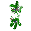
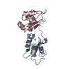
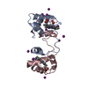
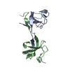
Hydrogenobacter thermophilus (bacteria)
MethodX-RAY DIFFRACTION / SYNCHROTRON / MOLECULAR REPLACEMENT / Resolution: 1.8 Å
AuthorsHayashi, Y. / Yamanaka, M. / Nagao, S. / Komori, H. / Higuchi, Y. / Hirota, S.
Funding support Japan, 2items
OrganizationGrant numberCountry
Japan Society for the Promotion of ScienceNo. 26288080 Japan
Japan Society for the Promotion of ScienceNo. 15K13744 Japan
CitationJournal: Sci Rep / Year: 2016
Title: Domain swapping oligomerization of thermostable c-type cytochrome in E. coli cells
Authors: Hayashi, Y. / Yamanaka, M. / Nagao, S. / Komori, H. / Higuchi, Y. / Hirota, S.
History
DepositionApr 28, 2015Deposition site: RCSB / Processing site: PDBJ
Revision 1.0Feb 10, 2016Provider: repository / Type: Initial release
Revision 1.1Feb 24, 2016Group: Database references
Revision 1.2Feb 19, 2020Group: Data collection / Derived calculations / Category: diffrn_source / pdbx_struct_oper_list
Item: _diffrn_source.pdbx_synchrotron_site / _pdbx_struct_oper_list.symmetry_operation
Revision 1.3Nov 8, 2023Group: Data collection / Database references / Refinement description
Category: chem_comp_atom / chem_comp_bond ...chem_comp_atom / chem_comp_bond / database_2 / pdbx_initial_refinement_model
Item: _database_2.pdbx_DOI / _database_2.pdbx_database_accession

-
Structure visualization

Structure viewerMolecule:
MolmilJmol/JSmol

Downloads & links

-
Assembly

Deposited unit
A: Cytochrome c-552
hetero molecules


Theoretical massNumber of molelcules
Total (without water)9,2052
Polymers8,5861
Non-polymers6191
Water52229
1
A: Cytochrome c-552
hetero molecules

A: Cytochrome c-552
hetero molecules


Theoretical massNumber of molelcules
Total (without water)18,4094
Polymers17,1722
Non-polymers1,2372
Water362
TypeNameSymmetry operationNumber
identity operation1_555x,y,z1
crystal symmetry operation4_465y-1,x+1,-z1
Buried area5440 Å2
ΔGint-64 kcal/mol
Surface area9290 Å2
MethodPISA
Unit cell
Length a, b, c (Å)45.808, 45.808, 78.304
Angle α, β, γ (deg.)90.00, 90.00, 120.00
Int Tables number152
Space group name H-MP3121

-
Components

#1: Protein Cytochrome c-552 / Cytochrome c552


Mass: 8586.015 Da / Num. of mol.: 1 / Fragment: UNP residues 19-98
Source method: isolated from a genetically manipulated source
Source: (gene. exp.) Hydrogenobacter thermophilus (strain DSM 6534 / IAM 12695 / TK-6) (bacteria)
Strain: DSM 6534 / IAM 12695 / TK-6 / Gene: HTH_0988, Hydth_0984 / Plasmid: pKO2 / Production host: ESCHERICHIA COLI (E. coli) / Strain (production host): JCB387 / References: UniProt: P15452
#2: Chemical ChemComp-HEC / HEME C / Heme C


Mass: 618.503 Da / Num. of mol.: 1 / Source method: obtained synthetically / Formula: C34H34FeN4O4
#3: Water ChemComp-HOH / water / Water


Mass: 18.015 Da / Num. of mol.: 29 / Source method: isolated from a natural source / Formula: H2O

-
Experimental details

-
Experiment

ExperimentMethod: X-RAY DIFFRACTION

-
Sample preparation

CrystalDensity Matthews: 2.76 Å3/Da / Density % sol: 55.47 %
Crystal growTemperature: 293 K / Method: vapor diffusion, sitting drop / pH: 6.5 / Details: 1.6 M sodium citrate buffer / Temp details: room temperature

-
Data collection

DiffractionMean temperature: 100 K
Diffraction sourceSource: SYNCHROTRON / Site: SPring-8 / Beamline: BL38B1 / Wavelength: 1 Å
DetectorType: ADSC QUANTUM 315 / Detector: CCD / Date: Jun 9, 2013
RadiationProtocol: SINGLE WAVELENGTH / Monochromatic (M) / Laue (L): M / Scattering type: x-ray
Radiation wavelengthWavelength: 1 Å / Relative weight: 1
ReflectionResolution: 1.8→50 Å / Num. obs: 9242 / % possible obs: 99.2 % / Observed criterion σ(F): 0 / Observed criterion σ(I): -3 / Redundancy: 9.7 % / Rmerge(I) obs: 0.07 / Net I/σ(I): 37.5
Reflection shellResolution: 1.8→1.86 Å / Redundancy: 7.1 % / Rmerge(I) obs: 0.649 / Mean I/σ(I) obs: 2.2 / % possible all: 93.9

-
Processing

Software
NameVersionClassification
REFMAC5.8.0107refinement
REFMAC5.8.0107data reduction
HKL-2000data scaling
MOLREPphasing
RefinementMethod to determine structure: MOLECULAR REPLACEMENT
Starting model: 3VYM
Resolution: 1.8→39.67 Å / Cor.coef. Fo:Fc: 0.972 / Cor.coef. Fo:Fc free: 0.955 / SU B: 3.982 / SU ML: 0.076 / Cross valid method: THROUGHOUT / ESU R: 0.103 / ESU R Free: 0.104 / Stereochemistry target values: MAXIMUM LIKELIHOOD / Details: HYDROGENS HAVE BEEN ADDED IN THE RIDING POSITIONS
RfactorNum. reflection% reflectionSelection details
Rfree0.2035 440 4.8 %RANDOM
Rwork0.16709 ---
obs0.1688 8721 98.72 %-
Solvent computationIon probe radii: 0.8 Å / Shrinkage radii: 0.8 Å / VDW probe radii: 1.2 Å / Solvent model: MASK
Displacement parametersBiso mean: 39.799 Å2
Baniso -1Baniso -2Baniso -3
1-1.37 Å20.69 Å20 Å2
2--1.37 Å2-0 Å2
3----4.45 Å2
Refinement stepCycle: 1 / Resolution: 1.8→39.67 Å
ProteinNucleic acidLigandSolventTotal
Num. atoms601 0 43 29 673
Refine LS restraints
Refine-IDTypeDev idealDev ideal targetNumber
X-RAY DIFFRACTIONr_bond_refined_d0.0320.02662
X-RAY DIFFRACTIONr_bond_other_d0.0060.02650
X-RAY DIFFRACTIONr_angle_refined_deg2.8472.176904
X-RAY DIFFRACTIONr_angle_other_deg1.27831501
X-RAY DIFFRACTIONr_dihedral_angle_1_deg6.136579
X-RAY DIFFRACTIONr_dihedral_angle_2_deg37.82926.1921
X-RAY DIFFRACTIONr_dihedral_angle_3_deg15.75715116
X-RAY DIFFRACTIONr_dihedral_angle_4_deg8.048151
X-RAY DIFFRACTIONr_chiral_restr0.1770.287
X-RAY DIFFRACTIONr_gen_planes_refined0.0190.021742
X-RAY DIFFRACTIONr_gen_planes_other0.0020.02125
X-RAY DIFFRACTIONr_nbd_refined
X-RAY DIFFRACTIONr_nbd_other
X-RAY DIFFRACTIONr_nbtor_refined
X-RAY DIFFRACTIONr_nbtor_other
X-RAY DIFFRACTIONr_xyhbond_nbd_refined
X-RAY DIFFRACTIONr_xyhbond_nbd_other
X-RAY DIFFRACTIONr_metal_ion_refined
X-RAY DIFFRACTIONr_metal_ion_other
X-RAY DIFFRACTIONr_symmetry_vdw_refined
X-RAY DIFFRACTIONr_symmetry_vdw_other
X-RAY DIFFRACTIONr_symmetry_hbond_refined
X-RAY DIFFRACTIONr_symmetry_hbond_other
X-RAY DIFFRACTIONr_symmetry_metal_ion_refined
X-RAY DIFFRACTIONr_symmetry_metal_ion_other
X-RAY DIFFRACTIONr_mcbond_it2.1072.11319
X-RAY DIFFRACTIONr_mcbond_other2.0922.104318
X-RAY DIFFRACTIONr_mcangle_it2.783.142397
X-RAY DIFFRACTIONr_mcangle_other2.7853.147398
X-RAY DIFFRACTIONr_scbond_it3.1132.427341
X-RAY DIFFRACTIONr_scbond_other3.0882.425337
X-RAY DIFFRACTIONr_scangle_it
X-RAY DIFFRACTIONr_scangle_other4.4763.512505
X-RAY DIFFRACTIONr_long_range_B_refined6.12718.303794
X-RAY DIFFRACTIONr_long_range_B_other6.13518.268793
X-RAY DIFFRACTIONr_rigid_bond_restr
X-RAY DIFFRACTIONr_sphericity_free
X-RAY DIFFRACTIONr_sphericity_bonded
LS refinement shellResolution: 1.799→1.846 Å / Total num. of bins used: 20
RfactorNum. reflection% reflection
Rfree0.292 30 -
Rwork0.256 586 -
obs--91.94 %
Refinement TLS params.

Method: refined / Refine-ID: X-RAY DIFFRACTION

IDL112)L122)L132)L222)L232)L332)S11 (Å °)S12 (Å °)S13 (Å °)S21 (Å °)S22 (Å °)S23 (Å °)S31 (Å °)S32 (Å °)S33 (Å °)T112)T122)T132)T222)T232)T332)Origin x (Å)Origin y (Å)Origin z (Å)
111.0560.1797-1.13260.84330.51984.44260.0904-0.6666-0.79680.08570.10170.21860.4272-0.1897-0.19210.0756-0.0326-0.00640.0910.09280.2188-51.113316.72674.4051
27.2283-1.0484-0.13496.9424-1.966310.1763-0.09150.14960.2014-0.10620.1350.2758-0.4208-0.4194-0.04350.1080.034-0.04150.0949-0.02560.1573-29.651915.5012-5.1844
30.05170.311-0.3482.857-6.233424.3935-0.05630.04690.0405-0.02080.0060.0342-1.40310.13430.05030.2151-0.0776-0.00770.23160.14260.4166-17.392518.009-11.3817
415.44061.04898.36968.9280.634433.0388-0.0861-0.04750.41410.0569-0.062-0.4791-0.47390.62920.14810.0749-0.01330.01110.0948-0.00060.1275-15.653711.2765-1.9889
52.5075-1.86091.98964.79121.10226.0555-0.12850.0020.02070.09940.1468-0.0726-0.08750.3892-0.01830.0703-0.03590.01940.07770.00150.1335-19.564111.02060.034
610.6222-0.4295-1.65798.2166-2.68899.74070.07790.5726-0.1079-0.3288-0.06870.28810.26010.0664-0.00920.1081-0.00410.01140.1223-0.04010.0636-25.10519.1245-12.4622
Refinement TLS group
IDRefine-IDRefine TLS-IDAuth asym-IDAuth seq-ID
1X-RAY DIFFRACTION1A1 - 18
2X-RAY DIFFRACTION2A19 - 33
3X-RAY DIFFRACTION3A34 - 42
4X-RAY DIFFRACTION4A43 - 48
5X-RAY DIFFRACTION5A49 - 70
6X-RAY DIFFRACTION6A71 - 80

+
About Yorodumi

-
News

-
Feb 9, 2022. New format data for meta-information of EMDB entries

New format data for meta-information of EMDB entries

  • Version 3 of the EMDB header file is now the official format.
  • The previous official version 1.9 will be removed from the archive.

Related info.:EMDB header

External links:wwPDB to switch to version 3 of the EMDB data model

-
Aug 12, 2020. Covid-19 info

Covid-19 info

URL: https://pdbj.org/emnavi/covid19.php

New page: Covid-19 featured information page in EM Navigator.

Related info.:Covid-19 info / Mar 5, 2020. Novel coronavirus structure data

+
Mar 5, 2020. Novel coronavirus structure data

Novel coronavirus structure data

Related info.:Yorodumi Speices / Aug 12, 2020. Covid-19 info

External links:COVID-19 featured content - PDBj / Molecule of the Month (242):Coronavirus Proteases

+
Jan 31, 2019. EMDB accession codes are about to change! (news from PDBe EMDB page)

EMDB accession codes are about to change! (news from PDBe EMDB page)

  • The allocation of 4 digits for EMDB accession codes will soon come to an end. Whilst these codes will remain in use, new EMDB accession codes will include an additional digit and will expand incrementally as the available range of codes is exhausted. The current 4-digit format prefixed with “EMD-” (i.e. EMD-XXXX) will advance to a 5-digit format (i.e. EMD-XXXXX), and so on. It is currently estimated that the 4-digit codes will be depleted around Spring 2019, at which point the 5-digit format will come into force.
  • The EM Navigator/Yorodumi systems omit the EMD- prefix.

Related info.:Q: What is EMD? / ID/Accession-code notation in Yorodumi/EM Navigator

External links:EMDB Accession Codes are Changing Soon! / Contact to PDBj

+
Jul 12, 2017. Major update of PDB

Major update of PDB

  • wwPDB released updated PDB data conforming to the new PDBx/mmCIF dictionary.
  • This is a major update changing the version number from 4 to 5, and with Remediation, in which all the entries are updated.
  • In this update, many items about electron microscopy experimental information are reorganized (e.g. em_software).
  • Now, EM Navigator and Yorodumi are based on the updated data.

External links:wwPDB Remediation / Enriched Model Files Conforming to OneDep Data Standards Now Available in the PDB FTP Archive

-
Yorodumi

Thousand views of thousand structures

  • Yorodumi is a browser for structure data from EMDB, PDB, SASBDB, etc.
  • This page is also the successor to EM Navigator detail page, and also detail information page/front-end page for Omokage search.
  • The word "yorodu" (or yorozu) is an old Japanese word meaning "ten thousand". "mi" (miru) is to see.

Related info.:EMDB / PDB / SASBDB / Comparison of 3 databanks / Yorodumi Search / Aug 31, 2016. New EM Navigator & Yorodumi / Yorodumi Papers / Jmol/JSmol / Function and homology information / Changes in new EM Navigator and Yorodumi

Read more