[English] 日本語
Yorodumi
- PDB-6h2j: Crystal structure of the HsdR subunit of the EcoR124I restriction... -

+
Open data


ID or keywords:

Loading...

-
Basic information

Entry
Database: PDB / ID: 6h2j
TitleCrystal structure of the HsdR subunit of the EcoR124I restriction enzyme with the C-terminal domain
ComponentsType I restriction enzyme R Protein
KeywordsHYDROLASE / EcoR124 / HsdR / C-terminal domain / restriction-modification enzyme / endonuclease / Escherichia coli
Function / homology
Function and homology information


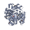
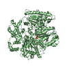
type I site-specific deoxyribonuclease / type I site-specific deoxyribonuclease activity / DNA restriction-modification system / DNA binding / ATP binding
Similarity search - Function
tt1808, chain A - #50 / Actin; Chain A, domain 4 - #50 / tt1808, chain A / Restriction endonuclease, type I, HsdR, N-terminal / Type I restriction enzyme R protein, C-terminal / SWI2/SNF2 ATPase / Type I restriction enzyme R protein N terminus (HSDR_N) / Type I restriction and modification enzyme - subunit R C terminal / SWI2/SNF2 ATPase / Restriction endonuclease, type I, HsdR ...tt1808, chain A - #50 / Actin; Chain A, domain 4 - #50 / tt1808, chain A / Restriction endonuclease, type I, HsdR, N-terminal / Type I restriction enzyme R protein, C-terminal / SWI2/SNF2 ATPase / Type I restriction enzyme R protein N terminus (HSDR_N) / Type I restriction and modification enzyme - subunit R C terminal / SWI2/SNF2 ATPase / Restriction endonuclease, type I, HsdR / Actin; Chain A, domain 4 / Superfamilies 1 and 2 helicase ATP-binding type-1 domain profile. / DEAD-like helicases superfamily / Helicase superfamily 1/2, ATP-binding domain / P-loop containing nucleotide triphosphate hydrolases / Alpha-Beta Complex / P-loop containing nucleoside triphosphate hydrolase / Rossmann fold / 3-Layer(aba) Sandwich / Alpha Beta
Similarity search - Domain/homology
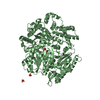
ADENOSINE-5'-TRIPHOSPHATE / Type I restriction enzyme EcoR124I/EcoR124II endonuclease subunit / Type I restriction enzyme EcoR124I/EcoR124II endonuclease subunit
Similarity search - Component
Biological speciesEscherichia coli (E. coli)
MethodX-RAY DIFFRACTION / SYNCHROTRON / MOLECULAR REPLACEMENT / Resolution: 2.6 Å
AuthorsGrinkevich, P. / Mesters, J.R. / Ettrich, R.H.
Funding support Czech Republic, 1items
OrganizationGrant numberCountry
Czech Science FoundationP207/12/2323 Czech Republic
CitationJournal: J. Biol. Chem. / Year: 2018
Title: Crystal structure of a novel domain of the motor subunit of the Type I restriction enzyme EcoR124 involved in complex assembly and DNA binding.
Authors: Grinkevich, P. / Sinha, D. / Iermak, I. / Guzanova, A. / Weiserova, M. / Ludwig, J. / Mesters, J.R. / Ettrich, R.H.
History
DepositionJul 13, 2018Deposition site: PDBE / Processing site: PDBE
Revision 1.0Aug 8, 2018Provider: repository / Type: Initial release
Revision 1.1Oct 10, 2018Group: Data collection / Database references / Category: citation
Item: _citation.journal_volume / _citation.page_first / _citation.page_last
Revision 1.2Jul 10, 2019Group: Data collection / Category: diffrn_source / Item: _diffrn_source.pdbx_synchrotron_site
Revision 1.3Jul 17, 2019Group: Data collection / Category: diffrn_source / Item: _diffrn_source.pdbx_synchrotron_site
Revision 1.4Jan 17, 2024Group: Data collection / Database references ...Data collection / Database references / Derived calculations / Refinement description
Category: chem_comp_atom / chem_comp_bond ...chem_comp_atom / chem_comp_bond / database_2 / pdbx_initial_refinement_model / struct_conn
Item: _database_2.pdbx_DOI / _database_2.pdbx_database_accession ..._database_2.pdbx_DOI / _database_2.pdbx_database_accession / _struct_conn.conn_type_id / _struct_conn.id / _struct_conn.pdbx_dist_value / _struct_conn.pdbx_leaving_atom_flag / _struct_conn.ptnr1_auth_asym_id / _struct_conn.ptnr1_auth_comp_id / _struct_conn.ptnr1_auth_seq_id / _struct_conn.ptnr1_label_asym_id / _struct_conn.ptnr1_label_atom_id / _struct_conn.ptnr1_label_comp_id / _struct_conn.ptnr1_label_seq_id / _struct_conn.ptnr2_auth_asym_id / _struct_conn.ptnr2_auth_comp_id / _struct_conn.ptnr2_auth_seq_id / _struct_conn.ptnr2_label_asym_id / _struct_conn.ptnr2_label_atom_id / _struct_conn.ptnr2_label_comp_id / _struct_conn.ptnr2_label_seq_id

-
Structure visualization

Structure viewerMolecule:
MolmilJmol/JSmol

Downloads & links

-
Assembly

Deposited unit
A: Type I restriction enzyme R Protein
B: Type I restriction enzyme R Protein
hetero molecules


Theoretical massNumber of molelcules
Total (without water)243,0286
Polymers241,9652
Non-polymers1,0634
Water6,900383
1
A: Type I restriction enzyme R Protein
hetero molecules


Theoretical massNumber of molelcules
Total (without water)121,5143
Polymers120,9821
Non-polymers5312
Water181
TypeNameSymmetry operationNumber
identity operation1_555x,y,z1
2
B: Type I restriction enzyme R Protein
hetero molecules


Theoretical massNumber of molelcules
Total (without water)121,5143
Polymers120,9821
Non-polymers5312
Water181
TypeNameSymmetry operationNumber
identity operation1_555x,y,z1
Unit cell
Length a, b, c (Å)123.910, 129.922, 161.010
Angle α, β, γ (deg.)90.00, 90.00, 90.00
Int Tables number19
Space group name H-MP212121

-
Components

#1: Protein Type I restriction enzyme R Protein


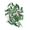
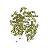
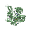
Mass: 120982.328 Da / Num. of mol.: 2
Source method: isolated from a genetically manipulated source
Source: (gene. exp.) Escherichia coli (E. coli) / Gene: hsdR / Production host: Escherichia coli (E. coli)
References: UniProt: Q304R3, UniProt: P10486*PLUS, type I site-specific deoxyribonuclease
#2: Chemical ChemComp-ATP / ADENOSINE-5'-TRIPHOSPHATE / Adenosine triphosphate


Mass: 507.181 Da / Num. of mol.: 2 / Source method: obtained synthetically / Formula: C10H16N5O13P3 / Comment: ATP, energy-carrying molecule*YM
#3: Chemical ChemComp-MG / MAGNESIUM ION


Mass: 24.305 Da / Num. of mol.: 2 / Source method: obtained synthetically / Formula: Mg
#4: Water ChemComp-HOH / water / Water


Mass: 18.015 Da / Num. of mol.: 383 / Source method: isolated from a natural source / Formula: H2O

-
Experimental details

-
Experiment

ExperimentMethod: X-RAY DIFFRACTION / Number of used crystals: 1

-
Sample preparation

CrystalDensity Matthews: 2.68 Å3/Da / Density % sol: 54.09 %
Crystal growTemperature: 277 K / Method: vapor diffusion, sitting drop
Details: 1 UL OF PROTEIN IN 20 MM PHOSPHATE PH 7.5, 100 MM KCL, 5 MM ATP WAS MIXED WITH 2 UL OF RESERVOIR, CONTAINING 0.2 M LI2SO4, 8 % PEG 20K, 8 % PEG 550 MME, 1.5 MM DTT

-
Data collection

DiffractionMean temperature: 100 K
Diffraction sourceSource: SYNCHROTRON / Site: EMBL/DESY, HAMBURG / Beamline: X12 / Wavelength: 0.987 Å
DetectorType: MARRESEARCH / Detector: CCD / Date: Oct 20, 2006
RadiationProtocol: MAD / Monochromatic (M) / Laue (L): M / Scattering type: x-ray
Radiation wavelengthWavelength: 0.987 Å / Relative weight: 1
ReflectionResolution: 2.6→20 Å / Num. obs: 80316 / % possible obs: 100 % / Redundancy: 9.4 % / Net I/σ(I): 18.2
Reflection shellResolution: 2.6→2.64 Å

-
Processing

Software
NameVersionClassification
PHENIX(1.13_2998: ???)refinement
DENZOdata reduction
SCALEPACKdata scaling
Auto-Rickshawphasing
RefinementMethod to determine structure: MOLECULAR REPLACEMENT
Starting model: 2w00, 5j3n
Resolution: 2.6→19.968 Å / SU ML: 0.33 / Cross valid method: FREE R-VALUE / σ(F): 1.33 / Phase error: 30.14 / Stereochemistry target values: ML
RfactorNum. reflection% reflection
Rfree0.2638 977 1.22 %
Rwork0.2027 --
obs0.2035 80202 99.93 %
Solvent computationShrinkage radii: 0.9 Å / VDW probe radii: 1.11 Å / Solvent model: FLAT BULK SOLVENT MODEL
Refinement stepCycle: LAST / Resolution: 2.6→19.968 Å
ProteinNucleic acidLigandSolventTotal
Num. atoms14917 0 64 383 15364
Refine LS restraints
Refine-IDTypeDev idealNumber
X-RAY DIFFRACTIONf_bond_d0.00915263
X-RAY DIFFRACTIONf_angle_d1.00820599
X-RAY DIFFRACTIONf_dihedral_angle_d5.4139216
X-RAY DIFFRACTIONf_chiral_restr0.0532262
X-RAY DIFFRACTIONf_plane_restr0.0062664
LS refinement shell
Resolution (Å)Rfactor RfreeNum. reflection RfreeRfactor RworkNum. reflection RworkRefine-ID% reflection obs (%)
2.6-2.73680.34911230.258611173X-RAY DIFFRACTION100
2.7368-2.90770.28741440.239511192X-RAY DIFFRACTION100
2.9077-3.13150.29341230.23111248X-RAY DIFFRACTION100
3.1315-3.44510.28721330.213911294X-RAY DIFFRACTION100
3.4451-3.94030.25961530.189711281X-RAY DIFFRACTION100
3.9403-4.95190.23511350.171611373X-RAY DIFFRACTION100
4.9519-19.96860.2421660.198711664X-RAY DIFFRACTION100
Refinement TLS params.

Method: refined / Refine-ID: X-RAY DIFFRACTION

IDL112)L122)L132)L222)L232)L332)S11 (Å °)S12 (Å °)S13 (Å °)S21 (Å °)S22 (Å °)S23 (Å °)S31 (Å °)S32 (Å °)S33 (Å °)T112)T122)T132)T222)T232)T332)Origin x (Å)Origin y (Å)Origin z (Å)
13.52520.70550.3580.9231-0.19260.9216-0.0268-0.11530.28460.0779-0.00470.0227-0.0177-0.03530.00110.30530.00590.01280.2832-0.01860.337733.77545.824630.086
21.1776-0.16540.06613.23311.04322.2384-0.03720.3031-0.2066-0.42360.0475-0.18030.08580.178-00.37780.00250.00860.4022-0.0090.292255.977628.551114.9611
32.75320.31820.59872.75020.1121.35060.2195-0.1174-0.50350.1448-0.0965-0.38910.36870.12030.0020.4549-0.0088-0.07840.32430.03990.442268.716119.69140.9436
42.3839-0.7611-0.59732.58841.12032.4956-0.0481-0.4716-0.04790.6876-0.0762-0.0121-0.1851-0.15730.00050.512-0.0848-0.07550.50650.02590.425466.298943.679855.5524
53.1728-0.78260.3340.24810.08070.97310.0023-0.0195-0.281-0.00420.0031-0.02790.13560.02230.0010.37010.0009-0.01730.3441-0.04050.289334.5835-12.695326.4665
63.14120.531-0.19511.71740.24660.2652-0.0728-0.3469-0.41960.28050.01210.04070.0977-0.07940.00380.35840.0384-0.03970.41450.01590.312224.84-14.998334.7004
71.6037-0.42460.24843.0788-0.41130.8634-0.13350.30010.1635-0.22980.12720.1343-0.1376-0.02480.00060.3251-0.0796-0.05810.4488-0.01620.26139.8380.979316.3236
81.35480.2519-1.15741.41980.33241.11570.0060.35730.1302-0.29430.05360.4217-0.0128-0.34220.00460.3304-0.0071-0.10180.39670.01880.4027-4.5191-4.292922.1447
93.1820.1942-0.51723.0382-0.37780.34270.1101-0.1830.57040.18020.04350.6611-0.4891-0.33810.00030.51980.040.11840.4044-0.03110.706-11.395513.357543.3995
101.31370.6025-0.49551.4291-0.98652.12630.12670.0060.19240.17110.00250.3472-0.12420.06270.00080.3252-0.00090.04240.3081-0.04330.3786-3.82010.89341.314
110.99460.57341.13671.833-0.55272.6135-0.104-0.55490.27610.7342-0.12660.24930.0015-0.22920.00090.6328-0.10480.09560.5963-0.02760.4465-3.6141-9.849661.4781
121.79240.50691.21250.15120.2061.5438-0.1355-0.44230.15540.52680.0625-0.56240.04760.5783-0.00091.0081-0.0862-0.1641.1017-0.03820.799725.8921-6.663467.1512
Refinement TLS group
IDRefine-IDRefine TLS-IDSelection details
1X-RAY DIFFRACTION1chain 'A' and (resid 13 through 259 )
2X-RAY DIFFRACTION2chain 'A' and (resid 260 through 468 )
3X-RAY DIFFRACTION3chain 'A' and (resid 469 through 699 )
4X-RAY DIFFRACTION4chain 'A' and (resid 700 through 885 )
5X-RAY DIFFRACTION5chain 'B' and (resid 13 through 122 )
6X-RAY DIFFRACTION6chain 'B' and (resid 123 through 225 )
7X-RAY DIFFRACTION7chain 'B' and (resid 226 through 425 )
8X-RAY DIFFRACTION8chain 'B' and (resid 426 through 483 )
9X-RAY DIFFRACTION9chain 'B' and (resid 484 through 605 )
10X-RAY DIFFRACTION10chain 'B' and (resid 606 through 771 )
11X-RAY DIFFRACTION11chain 'B' and (resid 772 through 859 )
12X-RAY DIFFRACTION12chain 'B' and (resid 860 through 1030 )

+
About Yorodumi

-
News

-
Feb 9, 2022. New format data for meta-information of EMDB entries

New format data for meta-information of EMDB entries

  • Version 3 of the EMDB header file is now the official format.
  • The previous official version 1.9 will be removed from the archive.

Related info.:EMDB header

External links:wwPDB to switch to version 3 of the EMDB data model

-
Aug 12, 2020. Covid-19 info

Covid-19 info

URL: https://pdbj.org/emnavi/covid19.php

New page: Covid-19 featured information page in EM Navigator.

Related info.:Covid-19 info / Mar 5, 2020. Novel coronavirus structure data

+
Mar 5, 2020. Novel coronavirus structure data

Novel coronavirus structure data

Related info.:Yorodumi Speices / Aug 12, 2020. Covid-19 info

External links:COVID-19 featured content - PDBj / Molecule of the Month (242):Coronavirus Proteases

+
Jan 31, 2019. EMDB accession codes are about to change! (news from PDBe EMDB page)

EMDB accession codes are about to change! (news from PDBe EMDB page)

  • The allocation of 4 digits for EMDB accession codes will soon come to an end. Whilst these codes will remain in use, new EMDB accession codes will include an additional digit and will expand incrementally as the available range of codes is exhausted. The current 4-digit format prefixed with “EMD-” (i.e. EMD-XXXX) will advance to a 5-digit format (i.e. EMD-XXXXX), and so on. It is currently estimated that the 4-digit codes will be depleted around Spring 2019, at which point the 5-digit format will come into force.
  • The EM Navigator/Yorodumi systems omit the EMD- prefix.

Related info.:Q: What is EMD? / ID/Accession-code notation in Yorodumi/EM Navigator

External links:EMDB Accession Codes are Changing Soon! / Contact to PDBj

+
Jul 12, 2017. Major update of PDB

Major update of PDB

  • wwPDB released updated PDB data conforming to the new PDBx/mmCIF dictionary.
  • This is a major update changing the version number from 4 to 5, and with Remediation, in which all the entries are updated.
  • In this update, many items about electron microscopy experimental information are reorganized (e.g. em_software).
  • Now, EM Navigator and Yorodumi are based on the updated data.

External links:wwPDB Remediation / Enriched Model Files Conforming to OneDep Data Standards Now Available in the PDB FTP Archive

-
Yorodumi

Thousand views of thousand structures

  • Yorodumi is a browser for structure data from EMDB, PDB, SASBDB, etc.
  • This page is also the successor to EM Navigator detail page, and also detail information page/front-end page for Omokage search.
  • The word "yorodu" (or yorozu) is an old Japanese word meaning "ten thousand". "mi" (miru) is to see.

Related info.:EMDB / PDB / SASBDB / Comparison of 3 databanks / Yorodumi Search / Aug 31, 2016. New EM Navigator & Yorodumi / Yorodumi Papers / Jmol/JSmol / Function and homology information / Changes in new EM Navigator and Yorodumi

Read more