 Movie
Movie Controller

[English] 日本語
 Yorodumi
- PDB-1mk4: Structure of Protein of Unknown Function YqjY from Bacillus subti... -
+ Open data

- Basic information
| Entry | Database: PDB / ID: 1mk4 | ||||||
|---|---|---|---|---|---|---|---|
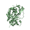
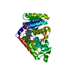
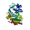
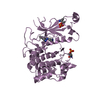
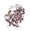
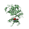
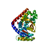
| Title | Structure of Protein of Unknown Function YqjY from Bacillus subtilis, Probable Acetyltransferase | ||||||
 Components Components | Hypothetical protein yqjY | ||||||
 Keywords Keywords | STRUCTURAL GENOMICS / UNKNOWN FUNCTION / alpha-beta-alpha sandwich / PSI / Protein Structure Initiative / Midwest Center for Structural Genomics / MCSG | ||||||
| Function / homology |  Function and homology information Function and homology informationacyltransferase activity, transferring groups other than amino-acyl groups Similarity search - Function | ||||||
| Biological species |  | ||||||
| Method |  X-RAY DIFFRACTION / SYNCHROTRON / MAD / Resolution: 1.7 Å X-RAY DIFFRACTION / SYNCHROTRON / MAD / Resolution: 1.7 Å | ||||||
 Authors Authors | Zhang, R. / Dementiva, I. / Mo, A. / Collart, F. / Joachimiak, A. / Midwest Center for Structural Genomics (MCSG) | ||||||
 Citation Citation | Journal: To be Published Title: 1.7A crystal structure of a hypothetical protein yqjY from Bacillus subtilis Authors: Zhang, R. / Dementiva, I. / Mo, A. / Collart, F. / Joachimiak, A. | ||||||
| History |
|
- Structure visualization
| Structure viewer | Molecule: MolmilJmol/JSmol |
|---|
- Downloads & links
-Download
| PDBx/mmCIF format | 1mk4.cif.gz | 81.2 KB | Display | PDBx/mmCIF format |
|---|---|---|---|---|
| PDB format | pdb1mk4.ent.gz | 61.5 KB | Display | PDB format |
| PDBx/mmJSON format | 1mk4.json.gz | Tree view | PDBx/mmJSON format | |
| Others |  Other downloads Other downloads |
-Validation report
| Arichive directory | https://data.pdbj.org/pub/pdb/validation_reports/mk/1mk4ftp://data.pdbj.org/pub/pdb/validation_reports/mk/1mk4 | HTTPS FTP |
|---|
-Related structure data
| Related structure data | |
|---|---|
| Similar structure data | |
| Other databases |


-Links
 PDBj
PDBj

- Assembly
| Deposited unit | 
| ||||||||
|---|---|---|---|---|---|---|---|---|---|
| 1 |
| ||||||||
| Unit cell |
| ||||||||
| Details | This protein existed as dimer(MolA and MolB) |
-Components
| #1: Protein | Mass: 18013.443 Da / Num. of mol.: 2 Source method: isolated from a genetically manipulated source Source: (gene. exp.) #2: Water | ChemComp-HOH / |  Mass: 18.015 Da / Num. of mol.: 325 / Source method: isolated from a natural source / Formula: H2O Mass: 18.015 Da / Num. of mol.: 325 / Source method: isolated from a natural source / Formula: H2O |
|---|
-Experimental details
-Experiment
| Experiment | Method: X-RAY DIFFRACTION / Number of used crystals: 1 |
|---|
- Sample preparation
| Crystal | Density Matthews: 3.1 Å3/Da / Density % sol: 60.3 % |
|---|---|
| Crystal grow | Temperature: 298 K / Method: vapor diffusion, hanging drop / pH: 7 Details: pH 7.0, VAPOR DIFFUSION, HANGING DROP, temperature 298K |
-Data collection
| Diffraction | Mean temperature: 100 K | ||||||||||||
|---|---|---|---|---|---|---|---|---|---|---|---|---|---|
| Diffraction source | Source: SYNCHROTRON / Site: APS  / Beamline: 19-ID / Wavelength: 0.9795,0.9798,0.94656 / Beamline: 19-ID / Wavelength: 0.9795,0.9798,0.94656 | ||||||||||||
| Detector | Type: SBC-2 / Detector: CCD / Date: Jul 31, 2002 / Details: mirrors | ||||||||||||
| Radiation | Monochromator: Si 111 Channel / Protocol: MAD / Monochromatic (M) / Laue (L): M / Scattering type: x-ray | ||||||||||||
| Radiation wavelength |
| ||||||||||||
| Reflection | Resolution: 1.7→28.09 Å / Num. all: 48445 / Num. obs: 48073 / % possible obs: 99.2 % / Observed criterion σ(F): 2 / Observed criterion σ(I): 2 / Redundancy: 7.14 % / Biso Wilson estimate: 15.5 Å2 / Rmerge(I) obs: 0.085 / Net I/σ(I): 27 | ||||||||||||
| Reflection shell | Resolution: 1.7→1.78 Å / Redundancy: 3.62 % / Rmerge(I) obs: 0.46 / Mean I/σ(I) obs: 4.59 / Num. unique all: 53174 / % possible all: 98.6 |
- Processing
| Software |
| ||||||||||||||||||||||||||||||||||||
|---|---|---|---|---|---|---|---|---|---|---|---|---|---|---|---|---|---|---|---|---|---|---|---|---|---|---|---|---|---|---|---|---|---|---|---|---|---|
| Refinement | Method to determine structure: MAD / Resolution: 1.7→28.09 Å / Rfactor Rfree error: 0.003 / Isotropic thermal model: RESTRAINED / Cross valid method: THROUGHOUT / σ(F): 0 / σ(I): 0 / Details: Freidel pairs were used for refinement.
| ||||||||||||||||||||||||||||||||||||
| Solvent computation | Solvent model: FLAT MODEL / Bsol: 41.4471 Å2 / ksol: 0.363244 e/Å3 | ||||||||||||||||||||||||||||||||||||
| Displacement parameters | Biso mean: 19.7 Å2
| ||||||||||||||||||||||||||||||||||||
| Refine analyze | Luzzati coordinate error free: 0.23 Å / Luzzati sigma a free: 0.17 Å | ||||||||||||||||||||||||||||||||||||
| Refinement step | Cycle: LAST / Resolution: 1.7→28.09 Å
| ||||||||||||||||||||||||||||||||||||
| Refine LS restraints |
| ||||||||||||||||||||||||||||||||||||
| LS refinement shell | Resolution: 1.7→1.81 Å / Rfactor Rfree error: 0.011 / Total num. of bins used: 6
| ||||||||||||||||||||||||||||||||||||
| Xplor file |
|
