 Movie
Movie Controller


+ Open data

- Basic information
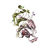
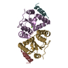
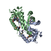
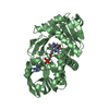
| Entry | Database: PDB / ID: 1i1d | ||||||
|---|---|---|---|---|---|---|---|
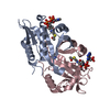
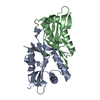
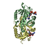
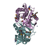
| Title | CRYSTAL STRUCTURE OF YEAST GNA1 BOUND TO COA AND GLNAC-6P | ||||||
 Components Components | GLUCOSAMINE-PHOSPHATE N-ACETYLTRANSFERASE | ||||||
 Keywords Keywords | TRANSFERASE / ALPHA/BETA / DOMAIN SWAPPING / GNAT CONSERVED CORE | ||||||
| Function / homology |  Function and homology information Function and homology informationSynthesis of UDP-N-acetyl-glucosamine / glucosamine-phosphate N-acetyltransferase / glucosamine 6-phosphate N-acetyltransferase activity / aralkylamine N-acetyltransferase activity / UDP-N-acetylglucosamine biosynthetic process / nucleus / cytoplasm Similarity search - Function | ||||||
| Biological species |  | ||||||
| Method |  X-RAY DIFFRACTION / SYNCHROTRON / MOLECULAR REPLACEMENT / Resolution: 1.8 Å X-RAY DIFFRACTION / SYNCHROTRON / MOLECULAR REPLACEMENT / Resolution: 1.8 Å | ||||||
 Authors Authors | Peneff, C. / Mengin-Lecreulx, D. / Bourne, Y. | ||||||
 Citation Citation | Journal: J.Biol.Chem. / Year: 2001 Title: The crystal structures of Apo and complexed Saccharomyces cerevisiae GNA1 shed light on the catalytic mechanism of an amino-sugar N-acetyltransferase. Authors: Peneff, C. / Mengin-Lecreulx, D. / Bourne, Y. | ||||||
| History |
|
- Structure visualization
| Structure viewer | Molecule: MolmilJmol/JSmol |
|---|
- Downloads & links
-Download
| PDBx/mmCIF format | 1i1d.cif.gz | 151.3 KB | Display | PDBx/mmCIF format |
|---|---|---|---|---|
| PDB format | pdb1i1d.ent.gz | 119.5 KB | Display | PDB format |
| PDBx/mmJSON format | 1i1d.json.gz | Tree view | PDBx/mmJSON format | |
| Others |  Other downloads Other downloads |
-Validation report
| Arichive directory | https://data.pdbj.org/pub/pdb/validation_reports/i1/1i1dftp://data.pdbj.org/pub/pdb/validation_reports/i1/1i1d | HTTPS FTP |
|---|
-Related structure data
| Related structure data |  1i12SC  1i21C S: Starting model for refinement C: citing same article ( |
|---|---|
| Similar structure data |


-Links
 PDBj
PDBj


- Assembly
| Deposited unit | 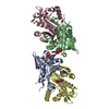
| |||||||||
|---|---|---|---|---|---|---|---|---|---|---|
| 1 | 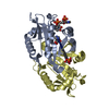
| |||||||||
| 2 | 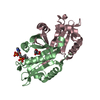
| |||||||||
| Unit cell |
| |||||||||
| Components on special symmetry positions |
|
-Components
| #1: Protein | Mass: 18316.074 Da / Num. of mol.: 4 / Mutation: S39C Source method: isolated from a genetically manipulated source Source: (gene. exp.) Gene: YFL017C / Plasmid: PQE30 / Production host:  References: UniProt: P43577, glucosamine-phosphate N-acetyltransferase #2: Sugar |   Type: D-saccharide, alpha linking / Mass: 301.188 Da / Num. of mol.: 3 Type: D-saccharide, alpha linking / Mass: 301.188 Da / Num. of mol.: 3Source method: isolated from a genetically manipulated source Formula: C8H16NO9P #3: Chemical |   Mass: 767.534 Da / Num. of mol.: 2 / Source method: obtained synthetically / Formula: C21H36N7O16P3S Mass: 767.534 Da / Num. of mol.: 2 / Source method: obtained synthetically / Formula: C21H36N7O16P3S#4: Chemical | ChemComp-IMD / |   Mass: 69.085 Da / Num. of mol.: 1 / Source method: obtained synthetically / Formula: C3H5N2 Mass: 69.085 Da / Num. of mol.: 1 / Source method: obtained synthetically / Formula: C3H5N2#5: Water | ChemComp-HOH / |  Mass: 18.015 Da / Num. of mol.: 542 / Source method: isolated from a natural source / Formula: H2O Mass: 18.015 Da / Num. of mol.: 542 / Source method: isolated from a natural source / Formula: H2O |
|---|
-Experimental details
-Experiment
| Experiment | Method: X-RAY DIFFRACTION / Number of used crystals: 1 |
|---|
- Sample preparation
| Crystal | Density Matthews: 2.4 Å3/Da / Density % sol: 48.65 % | ||||||||||||||||||||||||||||||||||||||||||||||||
|---|---|---|---|---|---|---|---|---|---|---|---|---|---|---|---|---|---|---|---|---|---|---|---|---|---|---|---|---|---|---|---|---|---|---|---|---|---|---|---|---|---|---|---|---|---|---|---|---|---|
| Crystal grow | Temperature: 293 K / Method: vapor diffusion, hanging drop / pH: 5.1 Details: PEG 600, IMIDAZOLE/MALATE, pH 5.10, VAPOR DIFFUSION, HANGING DROP, temperature 293K | ||||||||||||||||||||||||||||||||||||||||||||||||
| Crystal grow | *PLUS Temperature: 20 ℃ / pH: 8 | ||||||||||||||||||||||||||||||||||||||||||||||||
| Components of the solutions | *PLUS
|
-Data collection
| Diffraction | Mean temperature: 100 K | |||||||||
|---|---|---|---|---|---|---|---|---|---|---|
| Diffraction source | Source: SYNCHROTRON / Site: EMBL/DESY, HAMBURG  / Beamline: X11 / Wavelength: 1.1 / Wavelength: 1.1 Å / Beamline: X11 / Wavelength: 1.1 / Wavelength: 1.1 Å | |||||||||
| Detector | Type: MARRESEARCH / Detector: IMAGE PLATE / Date: Nov 27, 1999 | |||||||||
| Radiation | Protocol: SINGLE WAVELENGTH / Monochromatic (M) / Laue (L): M / Scattering type: x-ray | |||||||||
| Radiation wavelength |
| |||||||||
| Reflection | Resolution: 1.8→30 Å / Num. obs: 62765 / % possible obs: 97.4 % / Observed criterion σ(I): 2 / Redundancy: 2.2 % / Biso Wilson estimate: 18.4 Å2 / Rsym value: 4.8 / Net I/σ(I): 12.5 | |||||||||
| Reflection shell | Resolution: 1.8→1.86 Å / Redundancy: 2.2 % / Mean I/σ(I) obs: 1.8 / Rsym value: 41.2 / % possible all: 96.3 | |||||||||
| Reflection | *PLUS Highest resolution: 1.8 Å / Lowest resolution: 30 Å / Observed criterion σ(I): 2 / Redundancy: 2.2 % / Rmerge(I) obs: 0.048 | |||||||||
| Reflection shell | *PLUS % possible obs: 96.3 % / Rmerge(I) obs: 0.412 / Mean I/σ(I) obs: 1.8 |
- Processing
| Software |
| ||||||||||||||||||||
|---|---|---|---|---|---|---|---|---|---|---|---|---|---|---|---|---|---|---|---|---|---|
| Refinement | Method to determine structure: MOLECULAR REPLACEMENT Starting model: PDB ENTRY 1I12 Resolution: 1.8→24.12 Å / Rfactor Rfree error: 0.007 / Data cutoff high absF: 1131451.72 / Data cutoff low absF: 0 / Isotropic thermal model: RESTRAINED / Cross valid method: THROUGHOUT / σ(F): 0
| ||||||||||||||||||||
| Solvent computation | Solvent model: FLAT MODEL / Bsol: 54.22 Å2 / ksol: 0.376 e/Å3 | ||||||||||||||||||||
| Displacement parameters | Biso mean: 26.8 Å2
| ||||||||||||||||||||
| Refine analyze |
| ||||||||||||||||||||
| Refinement step | Cycle: LAST / Resolution: 1.8→24.12 Å
| ||||||||||||||||||||
| Refine LS restraints |
| ||||||||||||||||||||
| LS refinement shell | Resolution: 1.8→1.91 Å / Rfactor Rfree error: 0.023 / Total num. of bins used: 6
| ||||||||||||||||||||
| Xplor file |
| ||||||||||||||||||||
| Software | *PLUS Name: CNS / Version: 1 / Classification: refinement | ||||||||||||||||||||
| Refinement | *PLUS σ(F): 0 / % reflection Rfree: 1.8 % | ||||||||||||||||||||
| Solvent computation | *PLUS | ||||||||||||||||||||
| Displacement parameters | *PLUS Biso mean: 26.8 Å2 | ||||||||||||||||||||
| Refine LS restraints | *PLUS
| ||||||||||||||||||||
| LS refinement shell | *PLUS Rfactor Rfree: 0.302 / % reflection Rfree: 1.7 % / Rfactor Rwork: 0.259 |
