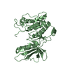
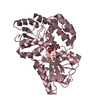
Journal: Cell Rep / Year: 2016 Title: A Molecular-Level Account of the Antigenic Hantaviral Surface. Authors: Sai Li / Ilona Rissanen / Antra Zeltina / Jussi Hepojoki / Jayna Raghwani / Karl Harlos / Oliver G Pybus / Juha T Huiskonen / Thomas A Bowden / Abstract: Hantaviruses, a geographically diverse group of zoonotic pathogens, initiate cell infection through the concerted action of Gn and Gc viral surface glycoproteins. Here, we describe the high- ...Hantaviruses, a geographically diverse group of zoonotic pathogens, initiate cell infection through the concerted action of Gn and Gc viral surface glycoproteins. Here, we describe the high-resolution crystal structure of the antigenic ectodomain of Gn from Puumala hantavirus (PUUV), a causative agent of hemorrhagic fever with renal syndrome. Fitting of PUUV Gn into an electron cryomicroscopy reconstruction of intact Gn-Gc spike complexes from the closely related but non-pathogenic Tula hantavirus localized Gn tetramers to the membrane-distal surface of the virion. The accuracy of the fitting was corroborated by epitope mapping and genetic analysis of available PUUV sequences. Interestingly, Gn exhibits greater non-synonymous sequence diversity than the less accessible Gc, supporting a role of the host humoral immune response in exerting selective pressure on the virus surface. The fold of PUUV Gn is likely to be widely conserved across hantaviruses.
Monochromator: DOUBLE CRYSTAL / Protocol: SINGLE WAVELENGTH / Monochromatic (M) / Laue (L): M / Scattering type: x-ray
Radiation wavelength
Wavelength: 0.9763 Å / Relative weight: 1
Reflection
Resolution: 2.28→62 Å / Num. obs: 43115 / % possible obs: 98.5 % / Observed criterion σ(I): -3 / Redundancy: 5.3 % / Rmerge(I) obs: 0.11 / Net I/σ(I): 12.1
Reflection shell
Resolution: 2.28→2.34 Å / Redundancy: 5 % / Rmerge(I) obs: 0.82 / Mean I/σ(I) obs: 2 / % possible all: 97.6
-
Processing
Software
Name
Version
Classification
REFMAC
5.8.0135
refinement
xia2
datareduction
xia2
datascaling
autoSHARP
phasing
Refinement
Method to determine structure: SAD Starting model: NONE Resolution: 2.28→73.26 Å / Cor.coef. Fo:Fc: 0.956 / Cor.coef. Fo:Fc free: 0.945 / SU B: 11.698 / SU ML: 0.161 / Cross valid method: THROUGHOUT / ESU R: 0.241 / ESU R Free: 0.189 / Stereochemistry target values: MAXIMUM LIKELIHOOD / Details: HYDROGENS HAVE BEEN ADDED IN THE RIDING POSITIONS.
Rfactor
Num. reflection
% reflection
Selection details
Rfree
0.21939
2171
5.1 %
RANDOM
Rwork
0.1886
-
-
-
obs
0.19015
40697
97.92 %
-
Solvent computation
Ion probe radii: 0.8 Å / Shrinkage radii: 0.8 Å / VDW probe radii: 1.2 Å / Solvent model: MASK
 Movie
Movie Controller


 Open data
 Basic information
 Components
 Keywords
 Function and homology information
 Puumala orthohantavirus
 X-RAY DIFFRACTION /
  Authors
 Citation

 Structure visualization
 Downloads & links
 Other downloads
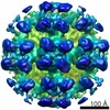


 PDBj
PDBj Assembly


 Homo sapiens (human) / References: UniProt: Q9WJ31
 Mass: 18.015 Da / Num. of mol.: 338 / Source method: isolated from a natural source / Formula: H2O
 Sample preparation
 Processing