 Movie
Movie Controller


+ Open data

- Basic information
| Entry | Database: PDB / ID: 5fyn | |||||||||
|---|---|---|---|---|---|---|---|---|---|---|
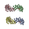
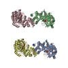
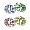
| Title | Sub-tomogram averaging of Tula virus glycoprotein spike | |||||||||
 Components Components | PUUMALA VIRUS GN GLYCOPROTEIN | |||||||||
 Keywords Keywords | VIRAL PROTEIN / TULA VIRUS / MEMBRANE PROTEIN / GLYCOPROTEIN / HANTAVIRUS / BUNYAVIRUS / RECEPTOR BINDING | |||||||||
| Function / homology |  Function and homology information Function and homology informationsymbiont-mediated suppression of host TRAF-mediated signal transduction / host cell Golgi membrane / host cell mitochondrion / host cell surface / symbiont-mediated suppression of host innate immune response / host cell endoplasmic reticulum membrane / fusion of virus membrane with host endosome membrane / symbiont entry into host cell / virion attachment to host cell / virion membrane ...symbiont-mediated suppression of host TRAF-mediated signal transduction / host cell Golgi membrane / host cell mitochondrion / host cell surface / symbiont-mediated suppression of host innate immune response / host cell endoplasmic reticulum membrane / fusion of virus membrane with host endosome membrane / symbiont entry into host cell / virion attachment to host cell / virion membrane / signal transduction / zinc ion binding Similarity search - Function | |||||||||
| Biological species |  PUUMALA VIRUS PUUMALA VIRUS | |||||||||
| Method | ELECTRON MICROSCOPY / electron tomography / cryo EM / Resolution: 15.6 Å | |||||||||
 Authors Authors | Li, S. / Rissanen, I. / Zeltina, A. / Hepojoki, J. / Raghwani, J. / Harlos, K. / Pybus, O.G. / Huiskonen, J.T. / Bowden, T.A. | |||||||||
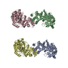
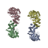
 Citation Citation | Journal: Cell Rep / Year: 2016 Title: A Molecular-Level Account of the Antigenic Hantaviral Surface. Authors: Sai Li / Ilona Rissanen / Antra Zeltina / Jussi Hepojoki / Jayna Raghwani / Karl Harlos / Oliver G Pybus / Juha T Huiskonen / Thomas A Bowden /   Abstract: Hantaviruses, a geographically diverse group of zoonotic pathogens, initiate cell infection through the concerted action of Gn and Gc viral surface glycoproteins. Here, we describe the high- ...Hantaviruses, a geographically diverse group of zoonotic pathogens, initiate cell infection through the concerted action of Gn and Gc viral surface glycoproteins. Here, we describe the high-resolution crystal structure of the antigenic ectodomain of Gn from Puumala hantavirus (PUUV), a causative agent of hemorrhagic fever with renal syndrome. Fitting of PUUV Gn into an electron cryomicroscopy reconstruction of intact Gn-Gc spike complexes from the closely related but non-pathogenic Tula hantavirus localized Gn tetramers to the membrane-distal surface of the virion. The accuracy of the fitting was corroborated by epitope mapping and genetic analysis of available PUUV sequences. Interestingly, Gn exhibits greater non-synonymous sequence diversity than the less accessible Gc, supporting a role of the host humoral immune response in exerting selective pressure on the virus surface. The fold of PUUV Gn is likely to be widely conserved across hantaviruses. | |||||||||
| History |
|
- Structure visualization
| Movie |
Movie viewer |
|---|---|
| Structure viewer | Molecule: MolmilJmol/JSmol |
- Downloads & links
-Download
| PDBx/mmCIF format | 5fyn.cif.gz | 141 KB | Display | PDBx/mmCIF format |
|---|---|---|---|---|
| PDB format | pdb5fyn.ent.gz | 109.2 KB | Display | PDB format |
| PDBx/mmJSON format | 5fyn.json.gz | Tree view | PDBx/mmJSON format | |
| Others |  Other downloads Other downloads |
-Validation report
| Arichive directory | https://data.pdbj.org/pub/pdb/validation_reports/fy/5fynftp://data.pdbj.org/pub/pdb/validation_reports/fy/5fyn | HTTPS FTP |
|---|
-Related structure data
| Related structure data | 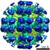 3364MC 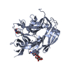 5fxuC M: map data used to model this data C: citing same article ( |
|---|---|
| Similar structure data |
-Links
 PDBj
PDBj- Assembly
| Deposited unit | 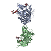
|
|---|---|
| 1 | 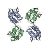
|
-Components
| #1: Protein | Mass: 38945.363 Da / Num. of mol.: 2 / Fragment: ECTODOMAIN, UNP RESIDUES 29-383 Source method: isolated from a genetically manipulated source Source: (gene. exp.) PUUMALA VIRUS / Plasmid: PHLSEC / Cell line (production host): HEK293 / Production host:  HOMO SAPIENS (human) / References: UniProt: Q9WJ31 HOMO SAPIENS (human) / References: UniProt: Q9WJ31#2: Polysaccharide | alpha-D-mannopyranose-(1-3)-beta-D-mannopyranose-(1-4)-2-acetamido-2-deoxy-beta-D-glucopyranose-(1- ...alpha-D-mannopyranose-(1-3)-beta-D-mannopyranose-(1-4)-2-acetamido-2-deoxy-beta-D-glucopyranose-(1-4)-2-acetamido-2-deoxy-beta-D-glucopyranose | Source method: isolated from a genetically manipulated source #3: Polysaccharide | Source method: isolated from a genetically manipulated source #4: Polysaccharide | beta-D-mannopyranose-(1-4)-2-acetamido-2-deoxy-beta-D-glucopyranose-(1-4)-2-acetamido-2-deoxy-beta- ...beta-D-mannopyranose-(1-4)-2-acetamido-2-deoxy-beta-D-glucopyranose-(1-4)-2-acetamido-2-deoxy-beta-D-glucopyranose | Source method: isolated from a genetically manipulated source Has protein modification | Y | |
|---|
-Experimental details
-Experiment
| Experiment | Method: ELECTRON MICROSCOPY |
|---|---|
| EM experiment | Aggregation state: PARTICLE / 3D reconstruction method: electron tomography |
- Sample preparation
| Component | Name: GLYCOPROTEIN SPIKE OF TULA HANTAVIRUS / Type: VIRUS |
|---|---|
| Buffer solution | Name: 25 MM TRIS, 75 MM NACL / pH: 8 / Details: 25 MM TRIS, 75 MM NACL |
| Specimen | Embedding applied: NO / Shadowing applied: NO / Staining applied: NO / Vitrification applied: YES |
| Specimen support | Details: HOLEY CARBON |
| Vitrification | Instrument: GATAN CRYOPLUNGE 3 / Cryogen name: ETHANE-PROPANE / Details: LIQUID ETHANE-PROPANE |
- Electron microscopy imaging
| Experimental equipment |  Model: Tecnai Polara / Image courtesy: FEI Company |
|---|---|
| Microscopy | Model: FEI POLARA 300 / Date: Aug 14, 2014 |
| Electron gun | Electron source:  FIELD EMISSION GUN / Accelerating voltage: 300 kV / Illumination mode: FLOOD BEAM FIELD EMISSION GUN / Accelerating voltage: 300 kV / Illumination mode: FLOOD BEAM |
| Electron lens | Mode: BRIGHT FIELD / Nominal magnification: 160000 X / Calibrated magnification: 37037 X / Nominal defocus max: 3800 nm / Nominal defocus min: 2000 nm / Cs: 2 mm |
| Specimen holder | Temperature: 100 K / Tilt angle max: 45 ° / Tilt angle min: -45 ° |
| Image recording | Electron dose: 60 e/Å2 / Film or detector model: GATAN K2 SUMMIT (4k x 4k) |
| Image scans | Num. digital images: 30 |
- Processing
| EM software |
| ||||||||||||||||||||
|---|---|---|---|---|---|---|---|---|---|---|---|---|---|---|---|---|---|---|---|---|---|
| CTF correction | Details: EACH TILTED IMAGE | ||||||||||||||||||||
| Symmetry | Point symmetry: C2 (2 fold cyclic) | ||||||||||||||||||||
| 3D reconstruction | Method: SUBTOMOGRAM AVERAGING / Resolution: 15.6 Å / Num. of particles: 5449 / Actual pixel size: 2.7 Å / Magnification calibration: KNOWN ATOMIC MODEL Details: SUBMISSION BASED ON EXPERIMENTAL DATA FROM EMDB EMD-3364. (DEPOSITION ID: 14310). Symmetry type: POINT | ||||||||||||||||||||
| Atomic model building | Protocol: OTHER / Space: REAL / Target criteria: Cross-correlation coefficient Details: METHOD--LOCAL CORRELATION REFINEMENT PROTOCOL--X-RAY | ||||||||||||||||||||
| Atomic model building | PDB-ID: 5FXU Accession code: 5FXU / Source name: PDB / Type: experimental model | ||||||||||||||||||||
| Refinement | Highest resolution: 15.6 Å | ||||||||||||||||||||
| Refinement step | Cycle: LAST / Highest resolution: 15.6 Å
|
