 Movie
Movie Controller


+ Open data

- Basic information
| Entry | Database: PDB / ID: 1o17 | |||||||||
|---|---|---|---|---|---|---|---|---|---|---|
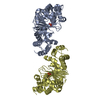
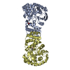
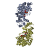
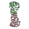
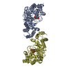
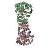
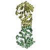
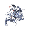
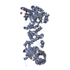
| Title | ANTHRANILATE PHOSPHORIBOSYL-TRANSFERASE (TRPD) | |||||||||
 Components Components | ANTHRANILATE PHOSPHORIBOSYLTRANSFERASE | |||||||||
 Keywords Keywords | TRANSFERASE / nucleoside-phosphorylases | |||||||||
| Function / homology |  Function and homology information Function and homology informationanthranilate phosphoribosyltransferase / anthranilate phosphoribosyltransferase activity / L-tryptophan biosynthetic process / magnesium ion binding / cytosol Similarity search - Function | |||||||||
| Biological species |   Sulfolobus solfataricus (archaea) Sulfolobus solfataricus (archaea) | |||||||||
| Method |  X-RAY DIFFRACTION / SYNCHROTRON / MAD / Resolution: 2.05 Å X-RAY DIFFRACTION / SYNCHROTRON / MAD / Resolution: 2.05 Å | |||||||||
 Authors Authors | Mayans, O. / Ivens, A. / Kirschner, K. / Wilmanns, M. | |||||||||
 Citation Citation | Journal: Embo J. / Year: 2002 Title: Structural analysis of two enzymes catalysing reverse metabolic reactions implies common ancestry Authors: Mayans, O. / Ivens, A. / Nissen, L. / Kirschner, K. / Wilmanns, M. #1: Journal: Eur.J.Biochem. / Year: 2001Title: Purification, characterization and crystallization of thermostable anthranilate phosphoribosyltransferase from Sulfolobus solfataricus Authors: Ivens, A. / Mayans, O. / Szadkowski, H. / Wilmanns, M. / Kirschner, K. | |||||||||
| History |
|
- Structure visualization
| Structure viewer | Molecule: MolmilJmol/JSmol |
|---|
- Downloads & links
-Download
| PDBx/mmCIF format | 1o17.cif.gz | 282.7 KB | Display | PDBx/mmCIF format |
|---|---|---|---|---|
| PDB format | pdb1o17.ent.gz | 230.7 KB | Display | PDB format |
| PDBx/mmJSON format | 1o17.json.gz | Tree view | PDBx/mmJSON format | |
| Others |  Other downloads Other downloads |
-Validation report
| Arichive directory | https://data.pdbj.org/pub/pdb/validation_reports/o1/1o17ftp://data.pdbj.org/pub/pdb/validation_reports/o1/1o17 | HTTPS FTP |
|---|
-Related structure data


-Links
 PDBj
PDBj- Assembly
| Deposited unit | 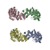
| ||||||||||
|---|---|---|---|---|---|---|---|---|---|---|---|
| 1 | 
| ||||||||||
| 2 | 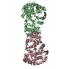
| ||||||||||
| Unit cell |
| ||||||||||
| Components on special symmetry positions |
| ||||||||||
| Details | biological active form: dimer |
-Components
| #1: Protein | Mass: 37619.516 Da / Num. of mol.: 4 Source method: isolated from a genetically manipulated source Source: (gene. exp.) Sulfolobus solfataricus (archaea) / Production host:  References: UniProt: P50384, anthranilate phosphoribosyltransferase #2: Water | ChemComp-HOH / |  Mass: 18.015 Da / Num. of mol.: 935 / Source method: isolated from a natural source / Formula: H2O Mass: 18.015 Da / Num. of mol.: 935 / Source method: isolated from a natural source / Formula: H2O |
|---|
-Experimental details
-Experiment
| Experiment | Method: X-RAY DIFFRACTION / Number of used crystals: 1 |
|---|
- Sample preparation
| Crystal | Density Matthews: 2.24 Å3/Da / Density % sol: 45.16 % | ||||||||||||||||||||||||
|---|---|---|---|---|---|---|---|---|---|---|---|---|---|---|---|---|---|---|---|---|---|---|---|---|---|
| Crystal grow | Temperature: 293 K / Method: vapor diffusion, hanging drop / pH: 6.5 Details: 0.1 M HEPES pH7.0 or 0.05 M MES pH6.0 & 12-18% PEG 1500, VAPOR DIFFUSION, HANGING DROP at 293K, pH 6.5 | ||||||||||||||||||||||||
| Crystal grow | *PLUS Temperature: 20 ℃ / pH: 7 / Details: Ivens, A., (2001) Eur.J.Biochem., 268, 2246. | ||||||||||||||||||||||||
| Components of the solutions | *PLUS
|
-Data collection
| Diffraction | Mean temperature: 100 K |
|---|---|
| Diffraction source | Source: SYNCHROTRON / Site: EMBL/DESY, HAMBURG  / Beamline: BW7B / Wavelength: 0.834 Å / Beamline: BW7B / Wavelength: 0.834 Å |
| Detector | Type: MARRESEARCH / Detector: IMAGE PLATE / Date: 1998 |
| Radiation | Protocol: SINGLE WAVELENGTH / Monochromatic (M) / Laue (L): M / Scattering type: x-ray |
| Radiation wavelength | Wavelength: 0.834 Å / Relative weight: 1 |
| Reflection | Resolution: 2.05→18 Å / Num. obs: 77715 / % possible obs: 92.7 % / Redundancy: 3.6 % / Biso Wilson estimate: 27.2 Å2 / Rsym value: 0.053 / Net I/σ(I): 10.8 |
| Reflection shell | Resolution: 2.05→2.09 Å / Redundancy: 1.9 % / Mean I/σ(I) obs: 1.8 / Num. unique all: 3382 / Rsym value: 0.245 / % possible all: 81.2 |
| Reflection | *PLUS Rmerge(I) obs: 0.053 |
| Reflection shell | *PLUS % possible obs: 81.2 % / Num. unique obs: 3382 / Rmerge(I) obs: 0.242 |
- Processing
| Software |
| ||||||||||||||||||||||||||||||||||||||||
|---|---|---|---|---|---|---|---|---|---|---|---|---|---|---|---|---|---|---|---|---|---|---|---|---|---|---|---|---|---|---|---|---|---|---|---|---|---|---|---|---|---|
| Refinement | Method to determine structure: MAD / Resolution: 2.05→18 Å / Cross valid method: THROUGHOUT / Stereochemistry target values: Engh & Huber
| ||||||||||||||||||||||||||||||||||||||||
| Displacement parameters |
| ||||||||||||||||||||||||||||||||||||||||
| Refinement step | Cycle: LAST / Resolution: 2.05→18 Å
| ||||||||||||||||||||||||||||||||||||||||
| Refine LS restraints |
| ||||||||||||||||||||||||||||||||||||||||
| Refinement | *PLUS Rfactor Rfree: 0.26 | ||||||||||||||||||||||||||||||||||||||||
| Solvent computation | *PLUS | ||||||||||||||||||||||||||||||||||||||||
| Displacement parameters | *PLUS | ||||||||||||||||||||||||||||||||||||||||
| Refine LS restraints | *PLUS
|
