 Movie
Movie Controller

[English] 日本語
 Yorodumi
- PDB-2gvq: Anthranilate phosphoribosyl-transferase (TRPD) from S. solfataric... -
+ Open data

- Basic information
| Entry | Database: PDB / ID: 2gvq | |||||||||
|---|---|---|---|---|---|---|---|---|---|---|
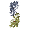
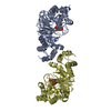
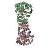
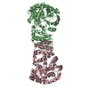
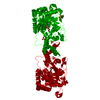
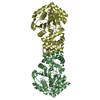
| Title | Anthranilate phosphoribosyl-transferase (TRPD) from S. solfataricus in complex with anthranilate | |||||||||
 Components Components | Anthranilate phosphoribosyltransferase | |||||||||
 Keywords Keywords | TRANSFERASE / PROTEIN-LIGAND COMPLEX | |||||||||
| Function / homology |  Function and homology information Function and homology informationanthranilate phosphoribosyltransferase / anthranilate phosphoribosyltransferase activity / L-tryptophan biosynthetic process / magnesium ion binding / cytosol Similarity search - Function | |||||||||
| Biological species |   Sulfolobus solfataricus (archaea) Sulfolobus solfataricus (archaea) | |||||||||
| Method |  X-RAY DIFFRACTION / SYNCHROTRON / FOURIER SYNTHESIS / Resolution: 2.43 Å X-RAY DIFFRACTION / SYNCHROTRON / FOURIER SYNTHESIS / Resolution: 2.43 Å | |||||||||
 Authors Authors | Marino, M. / Deuss, M. / Sterner, R. / Mayans, O. | |||||||||
 Citation Citation | Journal: J.Biol.Chem. / Year: 2006 Title: Structural and mutational analysis of substrate complexation by anthranilate phosphoribosyltransferase from Sulfolobus solfataricus. Authors: Marino, M. / Deuss, M. / Svergun, D.I. / Konarev, P.V. / Sterner, R. / Mayans, O. | |||||||||
| History |
|
- Structure visualization
| Structure viewer | Molecule: MolmilJmol/JSmol |
|---|
- Downloads & links
-Download
| PDBx/mmCIF format | 2gvq.cif.gz | 268.8 KB | Display | PDBx/mmCIF format |
|---|---|---|---|---|
| PDB format | pdb2gvq.ent.gz | 220 KB | Display | PDB format |
| PDBx/mmJSON format | 2gvq.json.gz | Tree view | PDBx/mmJSON format | |
| Others |  Other downloads Other downloads |
-Validation report
| Arichive directory | https://data.pdbj.org/pub/pdb/validation_reports/gv/2gvqftp://data.pdbj.org/pub/pdb/validation_reports/gv/2gvq | HTTPS FTP |
|---|
-Related structure data
| Related structure data |  1zxyC 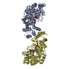 1zykC 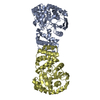 1o17S S: Starting model for refinement C: citing same article ( |
|---|---|
| Similar structure data |


-Links
 PDBj
PDBj- Assembly
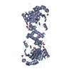
| Deposited unit | 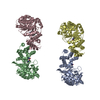
| ||||||||
|---|---|---|---|---|---|---|---|---|---|
| 1 | 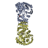
| ||||||||
| 2 | 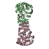
| ||||||||
| Unit cell |
| ||||||||
| Components on special symmetry positions |
|
-Components
| #1: Protein | Mass: 37619.516 Da / Num. of mol.: 4 Source method: isolated from a genetically manipulated source Source: (gene. exp.) Sulfolobus solfataricus (archaea) / Gene: TRPD / Plasmid: pQE40 / Production host:  References: UniProt: P50384, anthranilate phosphoribosyltransferase #2: Chemical | ChemComp-BE2 /   Type: L-peptide linking / Mass: 137.136 Da / Num. of mol.: 7 / Source method: obtained synthetically / Formula: C7H7NO2 Type: L-peptide linking / Mass: 137.136 Da / Num. of mol.: 7 / Source method: obtained synthetically / Formula: C7H7NO2#3: Water | ChemComp-HOH / |  Mass: 18.015 Da / Num. of mol.: 253 / Source method: isolated from a natural source / Formula: H2O Mass: 18.015 Da / Num. of mol.: 253 / Source method: isolated from a natural source / Formula: H2O |
|---|
-Experimental details
-Experiment
| Experiment | Method: X-RAY DIFFRACTION / Number of used crystals: 1 |
|---|
- Sample preparation
| Crystal | Density Matthews: 2.4 Å3/Da / Density % sol: 47.8 % |
|---|---|
| Crystal grow | Temperature: 293 K / Method: vapor diffusion, hanging drop / pH: 6 Details: PEG 1500, MES, pH 6.00, VAPOR DIFFUSION, HANGING DROP, temperature 293K |
-Data collection
| Diffraction | Mean temperature: 100 K |
|---|---|
| Diffraction source | Source: SYNCHROTRON / Site: SLS  / Beamline: X06SA / Wavelength: 0.951 / Wavelength: 0.951 Å / Beamline: X06SA / Wavelength: 0.951 / Wavelength: 0.951 Å |
| Detector | Type: MARRESEARCH / Detector: CCD / Date: Dec 11, 2002 |
| Radiation | Monochromator: SI (111) / Protocol: SINGLE WAVELENGTH / Monochromatic (M) / Laue (L): M / Scattering type: x-ray |
| Radiation wavelength | Wavelength: 0.951 Å / Relative weight: 1 |
| Reflection | Resolution: 2.43→20 Å / Num. obs: 49363 / % possible obs: 97.9 % / Redundancy: 3.84 % / Rsym value: 0.065 / Net I/σ(I): 10.99 |
| Reflection shell | Resolution: 2.43→2.5 Å / Redundancy: 3.41 % / Mean I/σ(I) obs: 3.1 / Rsym value: 0.43 / % possible all: 96.2 |
- Processing
| Software |
| ||||||||||||||||
|---|---|---|---|---|---|---|---|---|---|---|---|---|---|---|---|---|---|
| Refinement | Method to determine structure: FOURIER SYNTHESIS Starting model: PDB ENTRY 1O17 Resolution: 2.43→20 Å / Isotropic thermal model: ANISOTROPIC / σ(F): 0 / Stereochemistry target values: Engh & Huber
| ||||||||||||||||
| Displacement parameters | Biso mean: 42.39 Å2
| ||||||||||||||||
| Refinement step | Cycle: LAST / Resolution: 2.43→20 Å
| ||||||||||||||||
| Refine LS restraints |
|
