 Movie
Movie Controller

[English] 日本語
 Yorodumi
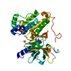
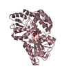
- PDB-3hpi: Crystal structure of maltose-binding protein mutant with bound sucrose -
+ Open data

- Basic information
| Entry | Database: PDB / ID: 3hpi | |||||||||
|---|---|---|---|---|---|---|---|---|---|---|
| Title | Crystal structure of maltose-binding protein mutant with bound sucrose | |||||||||
 Components Components | Maltose-binding periplasmic protein | |||||||||
 Keywords Keywords | SUGAR BINDING PROTEIN / periplasmic binding protein / MBP / Sugar transport / Transport | |||||||||
| Function / homology |  Function and homology information Function and homology informationdetection of maltose stimulus / maltose transport complex / carbohydrate transport / carbohydrate transmembrane transporter activity / maltose binding / maltose transport / maltodextrin transmembrane transport / ATP-binding cassette (ABC) transporter complex, substrate-binding subunit-containing / ATP-binding cassette (ABC) transporter complex / cell chemotaxis ...detection of maltose stimulus / maltose transport complex / carbohydrate transport / carbohydrate transmembrane transporter activity / maltose binding / maltose transport / maltodextrin transmembrane transport / ATP-binding cassette (ABC) transporter complex, substrate-binding subunit-containing / ATP-binding cassette (ABC) transporter complex / cell chemotaxis / outer membrane-bounded periplasmic space / periplasmic space / DNA damage response / membrane Similarity search - Function | |||||||||
| Biological species |  | |||||||||
| Method |  X-RAY DIFFRACTION / SYNCHROTRON / MOLECULAR REPLACEMENT / Resolution: 2 Å X-RAY DIFFRACTION / SYNCHROTRON / MOLECULAR REPLACEMENT / Resolution: 2 Å | |||||||||
 Authors Authors | Gould, A.D. / Shilton, B.H. | |||||||||
 Citation Citation | Journal: J.Biol.Chem. / Year: 2010 Title: Studies of the maltose transport system reveal a mechanism for coupling ATP hydrolysis to substrate translocation without direct recognition of substrate. Authors: Gould, A.D. / Shilton, B.H. #1: Journal: Proc.Natl.Acad.Sci.USA / Year: 2005 Title: Directed evolution of protein switches and their application to the creation of ligand-binding proteins. Authors: Guntas, G. / Mansell, T.J. / Kim, J.R. / Ostermeier, M. | |||||||||
| History |
|
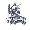
- Structure visualization
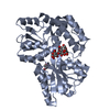
| Structure viewer | Molecule: MolmilJmol/JSmol |
|---|
- Downloads & links
-Download
| PDBx/mmCIF format | 3hpi.cif.gz | 159.9 KB | Display | PDBx/mmCIF format |
|---|---|---|---|---|
| PDB format | pdb3hpi.ent.gz | 123.8 KB | Display | PDB format |
| PDBx/mmJSON format | 3hpi.json.gz | Tree view | PDBx/mmJSON format | |
| Others |  Other downloads Other downloads |
-Validation report
| Arichive directory | https://data.pdbj.org/pub/pdb/validation_reports/hp/3hpiftp://data.pdbj.org/pub/pdb/validation_reports/hp/3hpi | HTTPS FTP |
|---|
-Related structure data
| Related structure data | 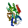 3kjtC  1anfS S: Starting model for refinement C: citing same article ( |
|---|---|
| Similar structure data |


-Links
 PDBj
PDBj


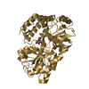
- Assembly
| Deposited unit | 
| ||||||||
|---|---|---|---|---|---|---|---|---|---|
| 1 | 
| ||||||||
| 2 | 
| ||||||||
| Unit cell |
|
-Components
| #1: Protein | Mass: 40908.371 Da / Num. of mol.: 2 / Mutation: D14L, K15F, W62Y, E111Y Source method: isolated from a genetically manipulated source Source: (gene. exp.) #2: Polysaccharide |   Source method: isolated from a genetically manipulated source Details: oligosaccharide with reducing-end-to-reducing-end glycosidic bond References: sucrose #3: Chemical | ChemComp-ZN /   Mass: 65.409 Da / Num. of mol.: 6 / Source method: obtained synthetically / Formula: Zn Mass: 65.409 Da / Num. of mol.: 6 / Source method: obtained synthetically / Formula: Zn#4: Chemical |   Mass: 59.044 Da / Num. of mol.: 2 / Source method: obtained synthetically / Formula: C2H3O2 Mass: 59.044 Da / Num. of mol.: 2 / Source method: obtained synthetically / Formula: C2H3O2#5: Water | ChemComp-HOH / |  Mass: 18.015 Da / Num. of mol.: 202 / Source method: isolated from a natural source / Formula: H2O Mass: 18.015 Da / Num. of mol.: 202 / Source method: isolated from a natural source / Formula: H2O |
|---|
-Experimental details
-Experiment
| Experiment | Method: X-RAY DIFFRACTION / Number of used crystals: 1 |
|---|
- Sample preparation
| Crystal | Density Matthews: 2.08 Å3/Da / Density % sol: 40.79 % |
|---|---|
| Crystal grow | Temperature: 289 K / Method: vapor diffusion / pH: 6.2 Details: PEG MME 5000, Sodium acetate, Sucrose, Magnesium chloride, Zinc chloride, pH 6.2, VAPOR DIFFUSION, temperature 289K |
-Data collection
| Diffraction | Mean temperature: 113 K |
|---|---|
| Diffraction source | Source: SYNCHROTRON / Site: CLSI  / Beamline: 08ID-1 / Wavelength: 0.9793 Å / Beamline: 08ID-1 / Wavelength: 0.9793 Å |
| Detector | Type: MARMOSAIC 225 mm CCD / Detector: CCD / Date: Mar 13, 2009 Details: DCM with cryo-cooled 1st crystal, sagitally bent 2nd crystal followed by vertically focusing mirror |
| Radiation | Monochromator: double crystal / Protocol: SINGLE WAVELENGTH / Scattering type: x-ray |
| Radiation wavelength | Wavelength: 0.9793 Å / Relative weight: 1 |
| Reflection | Resolution: 2→34.8 Å / Num. all: 44813 / Num. obs: 44813 / % possible obs: 96.2 % / Observed criterion σ(F): 0 / Observed criterion σ(I): 0 / Redundancy: 5.8 % / Biso Wilson estimate: 34 Å2 / Rmerge(I) obs: 0.092 / Χ2: 1.102 / Net I/σ(I): 16.124 |
| Reflection shell | Resolution: 2→2.07 Å / Redundancy: 4.4 % / Rmerge(I) obs: 0.468 / Mean I/σ(I) obs: 2.38 / Num. unique all: 4561 / Χ2: 1.019 / % possible all: 62.8 |
- Processing
| Software |
| ||||||||||||||||||||||||||||||||
|---|---|---|---|---|---|---|---|---|---|---|---|---|---|---|---|---|---|---|---|---|---|---|---|---|---|---|---|---|---|---|---|---|---|
| Refinement | Method to determine structure: MOLECULAR REPLACEMENT Starting model: PDB entry 1ANF Resolution: 2→34.8 Å / Occupancy max: 1 / Occupancy min: 1 / FOM work R set: 0.797 / Isotropic thermal model: anisotropic / Cross valid method: THROUGHOUT / σ(F): 2 / Stereochemistry target values: Engh & Huber
| ||||||||||||||||||||||||||||||||
| Solvent computation | Bsol: 51.209 Å2 | ||||||||||||||||||||||||||||||||
| Displacement parameters | Biso max: 80.7 Å2 / Biso mean: 32.554 Å2 / Biso min: 13.61 Å2
| ||||||||||||||||||||||||||||||||
| Refine analyze |
| ||||||||||||||||||||||||||||||||
| Refinement step | Cycle: LAST / Resolution: 2→34.8 Å
| ||||||||||||||||||||||||||||||||
| Refine LS restraints |
| ||||||||||||||||||||||||||||||||
| LS refinement shell | Resolution: 2→2.13 Å / Rfactor Rfree error: 0.021
| ||||||||||||||||||||||||||||||||
| Xplor file |
|
