 Movie
Movie Controller

[English] 日本語
 Yorodumi
- PDB-1mfl: The Structure of ERBIN PDZ domain bound to the Carboxy-terminal t... -
+ Open data

- Basic information
| Entry | Database: PDB / ID: 1mfl | ||||||
|---|---|---|---|---|---|---|---|
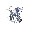
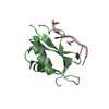
| Title | The Structure of ERBIN PDZ domain bound to the Carboxy-terminal tail of the ErbB2 Receptor | ||||||
 Components Components |
| ||||||
 Keywords Keywords | SIGNALING PROTEIN / PDZ DOMAIN / PHOSPHORYLATION / ERB-B2 / ERBIN | ||||||
| Function / homology |  Function and homology information Function and homology informationErbB-2 class receptor binding / basal protein localization / negative regulation of nucleotide-binding oligomerization domain containing 2 signaling pathway / hemidesmosome / negative regulation of immature T cell proliferation in thymus / ERBB3:ERBB2 complex / ERBB2-ERBB4 signaling pathway / postsynaptic specialization / immature T cell proliferation in thymus / GRB7 events in ERBB2 signaling ...ErbB-2 class receptor binding / basal protein localization / negative regulation of nucleotide-binding oligomerization domain containing 2 signaling pathway / hemidesmosome / negative regulation of immature T cell proliferation in thymus / ERBB3:ERBB2 complex / ERBB2-ERBB4 signaling pathway / postsynaptic specialization / immature T cell proliferation in thymus / GRB7 events in ERBB2 signaling / intermediate filament cytoskeleton organization / RNA polymerase I core binding / semaphorin receptor complex / negative regulation of monocyte chemotactic protein-1 production / Developmental Lineage of Mammary Stem Cells / establishment or maintenance of epithelial cell apical/basal polarity / ErbB-3 class receptor binding / Sema4D induced cell migration and growth-cone collapse / motor neuron axon guidance / regulation of microtubule-based process / : / PLCG1 events in ERBB2 signaling / RHOB GTPase cycle / enzyme-linked receptor protein signaling pathway / ERBB2 Activates PTK6 Signaling / ERBB2-EGFR signaling pathway / ERBB2-ERBB3 signaling pathway / neurotransmitter receptor localization to postsynaptic specialization membrane / Drug-mediated inhibition of ERBB2 signaling / Resistance of ERBB2 KD mutants to trastuzumab / Resistance of ERBB2 KD mutants to sapitinib / Resistance of ERBB2 KD mutants to tesevatinib / Resistance of ERBB2 KD mutants to neratinib / Resistance of ERBB2 KD mutants to osimertinib / Resistance of ERBB2 KD mutants to afatinib / Resistance of ERBB2 KD mutants to AEE788 / Resistance of ERBB2 KD mutants to lapatinib / Drug resistance in ERBB2 TMD/JMD mutants / RHOC GTPase cycle / positive regulation of MAP kinase activity / response to muramyl dipeptide / neuromuscular junction development / positive regulation of Rho protein signal transduction / positive regulation of transcription by RNA polymerase I / ERBB2 Regulates Cell Motility / Developmental Lineage of Mammary Gland Myoepithelial Cells / oligodendrocyte differentiation / semaphorin-plexin signaling pathway / basement membrane / PI3K events in ERBB2 signaling / RHOG GTPase cycle / RAC3 GTPase cycle / RHOA GTPase cycle / regulation of angiogenesis / RAC2 GTPase cycle / positive regulation of protein targeting to membrane / regulation of ERK1 and ERK2 cascade / Schwann cell development / regulation of postsynaptic membrane neurotransmitter receptor levels / coreceptor activity / protein targeting / Signaling by ERBB2 / TFAP2 (AP-2) family regulates transcription of growth factors and their receptors / peptidyl-tyrosine phosphorylation / RAC1 GTPase cycle / myelination / transmembrane receptor protein tyrosine kinase activity / GRB2 events in ERBB2 signaling / positive regulation of cell adhesion / SHC1 events in ERBB2 signaling / cell surface receptor protein tyrosine kinase signaling pathway / basal plasma membrane / cellular response to epidermal growth factor stimulus / Constitutive Signaling by Overexpressed ERBB2 / positive regulation of epithelial cell proliferation / Downregulation of ERBB2:ERBB3 signaling / integrin-mediated signaling pathway / positive regulation of translation / neuromuscular junction / wound healing / phosphatidylinositol 3-kinase/protein kinase B signal transduction / Signaling by ERBB2 TMD/JMD mutants / receptor protein-tyrosine kinase / Signaling by ERBB2 ECD mutants / Signaling by ERBB2 KD Mutants / receptor tyrosine kinase binding / structural constituent of cytoskeleton / cellular response to growth factor stimulus / cellular response to tumor necrosis factor / epidermal growth factor receptor signaling pathway / ruffle membrane / Downregulation of ERBB2 signaling / Constitutive Signaling by Aberrant PI3K in Cancer / neuron differentiation / cell junction / transmembrane signaling receptor activity / PIP3 activates AKT signaling / myelin sheath / heart development / PI5P, PP2A and IER3 Regulate PI3K/AKT Signaling Similarity search - Function | ||||||
| Biological species |  Homo sapiens (human) Homo sapiens (human) | ||||||
| Method |  X-RAY DIFFRACTION / MOLECULAR REPLACEMENT / Resolution: 1.88 Å X-RAY DIFFRACTION / MOLECULAR REPLACEMENT / Resolution: 1.88 Å | ||||||
 Authors Authors | Birrane, G. / Chung, J. / Ladias, J.A. | ||||||
 Citation Citation | Journal: J.Biol.Chem. / Year: 2003 Title: Novel mode of ligand recognition by the erbin PDZ domain Authors: Birrane, G. / Chung, J. / Ladias, J.A. #1: Journal: NAT.CELL BIOL. / Year: 2000Title: BIN: A BASOLATERAL PDZ PROTEIN THAT INTERACTS WITH THE MAMMALIAN ERBB2/HER2 RECEPTOR Authors: BORG, J.P. / MARCHETTO, S. / LEBIVIC, A. / OLLENDORFF, V. / JAULIN-BASTARD, F. / SAITO, H. / FOURNIER, E. / ADELAIDE, J. / MARGOLIS, B. / BIRNBAUM, D. #2: Journal: J.Biol.Chem. / Year: 2002Title: THE ERBIN PDZ DOMAIN BINDS WITH HIGH AFFINITY AND SPECIFICITY TO THE CARBOXYL TERMINI OF DELTA-CATENIN AND ARVCF Authors: LAURA, R.P. / WITT, A.S. / HELD, H.A. / GERSTNER, R. / DESHAYES, K. / KOEHLER, M.F. / KOSIK, K.S. / SIDHU, S.S. / LASKY, L.A. #3: Journal: J.Biol.Chem. / Year: 2001Title: ERBIN IS A PROTEIN CONCENTRATED AT POSTSYNAPTIC MEMBRANES THAT INTERACTS WITH PSD-95 Authors: HUANG, Y.Z. / WANG, Q. / XIONG, W.C. / MEI, L. #4: Journal: J.Biol.Chem. / Year: 2001Title: THE ERBB2/HER2 RECEPTOR DIFFERENTIALLY INTERACTS WITH ERBIN AND PICK1 PSD-95/DLG/ZO-1 DOMAIN PROTEINS Authors: JAULIN-BASTARD, F. / SAITO, H. / LEBIVIC, A. / OLLENDORFF, V. / MARCHETTO, S. / BIRNBAUM, D. / BORG, J.P. | ||||||
| History |
|
- Structure visualization
| Structure viewer | Molecule: MolmilJmol/JSmol |
|---|
- Downloads & links
-Download
| PDBx/mmCIF format | 1mfl.cif.gz | 33.6 KB | Display | PDBx/mmCIF format |
|---|---|---|---|---|
| PDB format | pdb1mfl.ent.gz | 22.4 KB | Display | PDB format |
| PDBx/mmJSON format | 1mfl.json.gz | Tree view | PDBx/mmJSON format | |
| Others |  Other downloads Other downloads |
-Validation report
| Arichive directory | https://data.pdbj.org/pub/pdb/validation_reports/mf/1mflftp://data.pdbj.org/pub/pdb/validation_reports/mf/1mfl | HTTPS FTP |
|---|
-Related structure data
| Related structure data |  1mfgSC S: Starting model for refinement C: citing same article ( |
|---|---|
| Similar structure data |

-Links
 PDBj
PDBj


- Assembly
| Deposited unit | 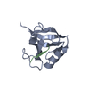
| ||||||||
|---|---|---|---|---|---|---|---|---|---|
| 1 |
| ||||||||
| Unit cell |
|
-Components
| #1: Protein | Mass: 10292.593 Da / Num. of mol.: 1 / Fragment: PDZ DOMAIN Source method: isolated from a genetically manipulated source Source: (gene. exp.) Homo sapiens (human) / Plasmid: PGEX-KT / Species (production host): Escherichia coli / Production host:  |
|---|---|
| #2: Protein/peptide | Mass: 1084.113 Da / Num. of mol.: 1 / Fragment: PEPTIDE LDVPV / Source method: obtained synthetically / Details: PEPTIDE SYNTHESIZED CHEMICALLY / References: UniProt: P04626*PLUS |
| #3: Water | ChemComp-HOH /  Mass: 18.015 Da / Num. of mol.: 72 / Source method: isolated from a natural source / Formula: H2O Mass: 18.015 Da / Num. of mol.: 72 / Source method: isolated from a natural source / Formula: H2O |
| Has protein modification | N |
-Experimental details
-Experiment
| Experiment | Method: X-RAY DIFFRACTION / Number of used crystals: 1 |
|---|
- Sample preparation
| Crystal | Density Matthews: 2.03 Å3/Da / Density % sol: 39.38 % | ||||||||||||||||||||||||||||||||||||||||||||||||||||||||
|---|---|---|---|---|---|---|---|---|---|---|---|---|---|---|---|---|---|---|---|---|---|---|---|---|---|---|---|---|---|---|---|---|---|---|---|---|---|---|---|---|---|---|---|---|---|---|---|---|---|---|---|---|---|---|---|---|---|
| Crystal grow | Temperature: 292 K / Method: vapor diffusion, sitting drop / pH: 4.5 Details: 23-26% PEG 4000, 20% GLYCEROL, 100mM AMMONIUM ACETATE, 100mM SODIUM ACETATE, pH 4.5, VAPOR DIFFUSION, SITTING DROP, temperature 292K | ||||||||||||||||||||||||||||||||||||||||||||||||||||||||
| Crystal grow | *PLUS Temperature: 20 ℃ / pH: 8.3 | ||||||||||||||||||||||||||||||||||||||||||||||||||||||||
| Components of the solutions | *PLUS
|
-Data collection
| Diffraction | Mean temperature: 100 K |
|---|---|
| Diffraction source | Source: ROTATING ANODE / Type: RIGAKU RU300 / Wavelength: 1.5418 Å |
| Detector | Type: RIGAKU RAXIS IV / Detector: IMAGE PLATE / Date: Jun 4, 2002 / Details: MIRRORS |
| Radiation | Monochromator: NI FILTER / Protocol: SINGLE WAVELENGTH / Monochromatic (M) / Laue (L): M / Scattering type: x-ray |
| Radiation wavelength | Wavelength: 1.5418 Å / Relative weight: 1 |
| Reflection | Resolution: 1.88→30.6 Å / Num. all: 7196 / Num. obs: 7068 / % possible obs: 95 % / Observed criterion σ(F): 0 / Observed criterion σ(I): -3 / Redundancy: 3.73 % / Biso Wilson estimate: 13 Å2 / Rmerge(I) obs: 0.028 / Rsym value: 0.028 / Net I/σ(I): 44.7 |
| Reflection shell | Resolution: 1.88→1.95 Å / Redundancy: 3.34 % / Rmerge(I) obs: 0.096 / Mean I/σ(I) obs: 13.3 / Num. unique all: 660 / Rsym value: 0.096 / % possible all: 90.2 |
| Reflection shell | *PLUS Lowest resolution: 1.93 Å / % possible obs: 90.2 % |
- Processing
| Software |
| ||||||||||||||||||||||||||||||||||||||||||||||||||||||||||||||||||||||||||||||||||||||||||||||||||||
|---|---|---|---|---|---|---|---|---|---|---|---|---|---|---|---|---|---|---|---|---|---|---|---|---|---|---|---|---|---|---|---|---|---|---|---|---|---|---|---|---|---|---|---|---|---|---|---|---|---|---|---|---|---|---|---|---|---|---|---|---|---|---|---|---|---|---|---|---|---|---|---|---|---|---|---|---|---|---|---|---|---|---|---|---|---|---|---|---|---|---|---|---|---|---|---|---|---|---|---|---|---|
| Refinement | Method to determine structure: MOLECULAR REPLACEMENT Starting model: PDB ENTRY 1MFG Resolution: 1.88→30.57 Å / Cor.coef. Fo:Fc: 0.952 / Cor.coef. Fo:Fc free: 0.941 / Cross valid method: THROUGHOUT / σ(F): 0 / σ(I): 0 / Stereochemistry target values: MAXIMUM LIKELIHOOD / Details: HYDROGENS HAVE BEEN ADDED IN THE RIDING POSITIONS
| ||||||||||||||||||||||||||||||||||||||||||||||||||||||||||||||||||||||||||||||||||||||||||||||||||||
| Solvent computation | Ion probe radii: 0.8 Å / Shrinkage radii: 0.8 Å / VDW probe radii: 1.4 Å / Solvent model: BABINET MODEL WITH MASK | ||||||||||||||||||||||||||||||||||||||||||||||||||||||||||||||||||||||||||||||||||||||||||||||||||||
| Displacement parameters | Biso mean: 18.168 Å2
| ||||||||||||||||||||||||||||||||||||||||||||||||||||||||||||||||||||||||||||||||||||||||||||||||||||
| Refinement step | Cycle: LAST / Resolution: 1.88→30.57 Å
| ||||||||||||||||||||||||||||||||||||||||||||||||||||||||||||||||||||||||||||||||||||||||||||||||||||
| Refine LS restraints |
| ||||||||||||||||||||||||||||||||||||||||||||||||||||||||||||||||||||||||||||||||||||||||||||||||||||
| LS refinement shell | Resolution: 1.88→1.929 Å / Total num. of bins used: 20 /
| ||||||||||||||||||||||||||||||||||||||||||||||||||||||||||||||||||||||||||||||||||||||||||||||||||||
| Refinement | *PLUS Lowest resolution: 30.6 Å / Num. reflection obs: 6737 / % reflection Rfree: 5 % / Rfactor Rfree: 0.216 / Rfactor Rwork: 0.167 | ||||||||||||||||||||||||||||||||||||||||||||||||||||||||||||||||||||||||||||||||||||||||||||||||||||
| Solvent computation | *PLUS | ||||||||||||||||||||||||||||||||||||||||||||||||||||||||||||||||||||||||||||||||||||||||||||||||||||
| Displacement parameters | *PLUS |
