 Movie
Movie Controller

[English] 日本語
 Yorodumi
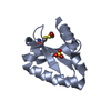
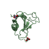
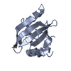
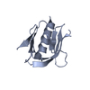
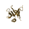
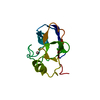
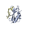
- PDB-4i67: Crystal structure of the RRM domain of RNA helicase HERA from T. ... -
+ Open data

- Basic information
| Entry | Database: PDB / ID: 4i67 | ||||||
|---|---|---|---|---|---|---|---|
| Title | Crystal structure of the RRM domain of RNA helicase HERA from T. thermophilus in complex with GGGC RNA | ||||||
 Components Components |
| ||||||
 Keywords Keywords | HYDROLASE/RNA / unwinding / ATPase / heat resistant / RNA recognition motif / RNA binding / DEAD box protein / HYDROLASE-RNA complex | ||||||
| Function / homology |  Function and homology information Function and homology informationnucleic acid binding / RNA helicase activity / hydrolase activity / ATP binding / metal ion binding / cytosol Similarity search - Function | ||||||
| Biological species |   Thermus thermophilus (bacteria) Thermus thermophilus (bacteria) | ||||||
| Method |  X-RAY DIFFRACTION / SYNCHROTRON / MOLECULAR REPLACEMENT / Resolution: 2.33 Å X-RAY DIFFRACTION / SYNCHROTRON / MOLECULAR REPLACEMENT / Resolution: 2.33 Å | ||||||
 Authors Authors | Rudolph, M.G. / Klostermeier, D. | ||||||
 Citation Citation | Journal: Nucleic Acids Res. / Year: 2013 Title: Recognition of two distinct elements in the RNA substrate by the RNA-binding domain of the T. thermophilus DEAD box helicase Hera. Authors: Steimer, L. / Wurm, J.P. / Linden, M.H. / Rudolph, M.G. / Wohnert, J. / Klostermeier, D. | ||||||
| History |
|
- Structure visualization
| Structure viewer | Molecule: MolmilJmol/JSmol |
|---|
- Downloads & links
-Download
| PDBx/mmCIF format | 4i67.cif.gz | 49.1 KB | Display | PDBx/mmCIF format |
|---|---|---|---|---|
| PDB format | pdb4i67.ent.gz | 34.6 KB | Display | PDB format |
| PDBx/mmJSON format | 4i67.json.gz | Tree view | PDBx/mmJSON format | |
| Others |  Other downloads Other downloads |
-Validation report
| Arichive directory | https://data.pdbj.org/pub/pdb/validation_reports/i6/4i67ftp://data.pdbj.org/pub/pdb/validation_reports/i6/4i67 | HTTPS FTP |
|---|
-Related structure data
| Related structure data |  4i68C  4i69C  3i31S S: Starting model for refinement C: citing same article ( |
|---|---|
| Similar structure data |


-Links
 PDBj
PDBj- Assembly
| Deposited unit | 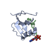
| ||||||||
|---|---|---|---|---|---|---|---|---|---|
| 1 |
| ||||||||
| Unit cell |
|
-Components
| #1: Protein | Mass: 9578.012 Da / Num. of mol.: 1 / Fragment: RRM domain (UNP residues 431-517) Source method: isolated from a genetically manipulated source Source: (gene. exp.) Thermus thermophilus (bacteria) / Gene: TT_C1895 / Production host: |
|---|---|
| #2: RNA chain | Mass: 1375.821 Da / Num. of mol.: 1 / Source method: obtained synthetically / Details: 23S ribosomal RNA fragment |
| #3: Water | ChemComp-HOH /  Mass: 18.015 Da / Num. of mol.: 11 / Source method: isolated from a natural source / Formula: H2O Mass: 18.015 Da / Num. of mol.: 11 / Source method: isolated from a natural source / Formula: H2O |
-Experimental details
-Experiment
| Experiment | Method: X-RAY DIFFRACTION / Number of used crystals: 1 |
|---|
- Sample preparation
| Crystal | Density Matthews: 2.98 Å3/Da / Density % sol: 58.76 % |
|---|---|
| Crystal grow | Temperature: 298 K / Method: vapor diffusion, sitting drop Details: 1.06 M sodium malonate, pH 6.0, 0.1 M Tris-HCl, pH 7.5, 0.13 M potassium/sodium phosphate, VAPOR DIFFUSION, SITTING DROP, temperature 298K PH range: 6-7.5 |
-Data collection
| Diffraction | Mean temperature: 100 K | |||||||||
|---|---|---|---|---|---|---|---|---|---|---|
| Diffraction source | Source: SYNCHROTRON / Site: SLS  / Beamline: X10SA / Wavelength: 0.99997 / Wavelength: 1 Å / Beamline: X10SA / Wavelength: 0.99997 / Wavelength: 1 Å | |||||||||
| Detector | Type: DECTRIS PILATUS 6M / Detector: PIXEL / Date: Jun 21, 2012 | |||||||||
| Radiation | Monochromator: double crystal Si(111) / Protocol: SINGLE WAVELENGTH / Monochromatic (M) / Laue (L): M / Scattering type: x-ray | |||||||||
| Radiation wavelength |
| |||||||||
| Reflection | Resolution: 2.33→44.2 Å / Num. obs: 5963 / % possible obs: 99.9 % / Observed criterion σ(F): 0 / Observed criterion σ(I): 0 / Redundancy: 9.76 % / Rmerge(I) obs: 0.052 / Rsym value: 0.052 / Net I/σ(I): 19.1 | |||||||||
| Reflection shell | Resolution: 2.33→2.43 Å / Redundancy: 9.9 % / Rmerge(I) obs: 0.868 / Mean I/σ(I) obs: 1.2 / Rsym value: 0.868 / % possible all: 99.4 |
- Processing
| Software |
| |||||||||||||||||||||||||||||||||||||||||||||||||||||||||||||||||||||||||||
|---|---|---|---|---|---|---|---|---|---|---|---|---|---|---|---|---|---|---|---|---|---|---|---|---|---|---|---|---|---|---|---|---|---|---|---|---|---|---|---|---|---|---|---|---|---|---|---|---|---|---|---|---|---|---|---|---|---|---|---|---|---|---|---|---|---|---|---|---|---|---|---|---|---|---|---|---|
| Refinement | Method to determine structure: MOLECULAR REPLACEMENT Starting model: PDB ENTRY 3I31 Resolution: 2.33→38.293 Å / SU ML: 0.34 / σ(F): 1.38 / Phase error: 32.49 / Stereochemistry target values: ML
| |||||||||||||||||||||||||||||||||||||||||||||||||||||||||||||||||||||||||||
| Solvent computation | Shrinkage radii: 0.9 Å / VDW probe radii: 1.11 Å / Solvent model: FLAT BULK SOLVENT MODEL | |||||||||||||||||||||||||||||||||||||||||||||||||||||||||||||||||||||||||||
| Refinement step | Cycle: LAST / Resolution: 2.33→38.293 Å
| |||||||||||||||||||||||||||||||||||||||||||||||||||||||||||||||||||||||||||
| Refine LS restraints |
| |||||||||||||||||||||||||||||||||||||||||||||||||||||||||||||||||||||||||||
| LS refinement shell |
| |||||||||||||||||||||||||||||||||||||||||||||||||||||||||||||||||||||||||||
| Refinement TLS params. | Method: refined / Refine-ID: X-RAY DIFFRACTION
| |||||||||||||||||||||||||||||||||||||||||||||||||||||||||||||||||||||||||||
| Refinement TLS group |
|
