 Movie
Movie Controller

[English] 日本語
 Yorodumi
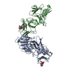
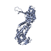
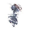
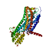
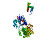
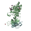
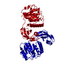
- PDB-1mdm: INHIBITED FRAGMENT OF ETS-1 AND PAIRED DOMAIN OF PAX5 BOUND TO DNA -
+ Open data

- Basic information
| Entry | Database: PDB / ID: 1mdm | ||||||
|---|---|---|---|---|---|---|---|
| Title | INHIBITED FRAGMENT OF ETS-1 AND PAIRED DOMAIN OF PAX5 BOUND TO DNA | ||||||
 Components Components |
| ||||||
 Keywords Keywords | TRANSCRIPTION/DNA / Transcription factor / ternary complex / autoinhibition / ETS domain / Paired domain / TRANSCRIPTION-DNA COMPLEX | ||||||
| Function / homology |  Function and homology information Function and homology informationRUNX1 regulates transcription of genes involved in BCR signaling / Oncogene Induced Senescence / regulation of extracellular matrix disassembly / PML body organization / sensory organ development / positive regulation of leukocyte adhesion to vascular endothelial cell / histone acetyltransferase binding / negative regulation of cell cycle / regulation of angiogenesis / positive regulation of blood vessel endothelial cell migration ...RUNX1 regulates transcription of genes involved in BCR signaling / Oncogene Induced Senescence / regulation of extracellular matrix disassembly / PML body organization / sensory organ development / positive regulation of leukocyte adhesion to vascular endothelial cell / histone acetyltransferase binding / negative regulation of cell cycle / regulation of angiogenesis / positive regulation of blood vessel endothelial cell migration / immune system process / positive regulation of endothelial cell migration / positive regulation of erythrocyte differentiation / B cell differentiation / transcription corepressor binding / cell motility / positive regulation of miRNA transcription / negative regulation of inflammatory response / positive regulation of angiogenesis / sequence-specific double-stranded DNA binding / positive regulation of inflammatory response / nervous system development / DNA-binding transcription activator activity, RNA polymerase II-specific / spermatogenesis / transcription regulator complex / regulation of apoptotic process / transcription by RNA polymerase II / sequence-specific DNA binding / DNA-binding transcription factor binding / nucleic acid binding / RNA polymerase II-specific DNA-binding transcription factor binding / DNA-binding transcription factor activity, RNA polymerase II-specific / cell differentiation / transcription cis-regulatory region binding / positive regulation of cell migration / RNA polymerase II cis-regulatory region sequence-specific DNA binding / DNA-binding transcription factor activity / response to antibiotic / positive regulation of cell population proliferation / positive regulation of gene expression / regulation of transcription by RNA polymerase II / positive regulation of DNA-templated transcription / chromatin / positive regulation of transcription by RNA polymerase II / DNA binding / nucleoplasm / identical protein binding / nucleus / cytoplasm Similarity search - Function | ||||||
| Biological species |  Homo sapiens (human) Homo sapiens (human) | ||||||
| Method |  X-RAY DIFFRACTION / MOLECULAR REPLACEMENT / Resolution: 2.8 Å X-RAY DIFFRACTION / MOLECULAR REPLACEMENT / Resolution: 2.8 Å | ||||||
 Authors Authors | Garvie, C.W. / Pufall, M.A. / Graves, B.J. / Wolberger, C. | ||||||
 Citation Citation | Journal: J.Biol.Chem. / Year: 2002 Title: STRUCTURAL ANALYSIS OF THE AUTOINHIBITION OF ETS-1 AND ITS ROLE IN PROTEIN PARTNERSHIPS Authors: Garvie, C.W. / Pufall, M.A. / Graves, B.J. / Wolberger, C. | ||||||
| History |
|
- Structure visualization
| Structure viewer | Molecule: MolmilJmol/JSmol |
|---|
- Downloads & links
-Download
| PDBx/mmCIF format | 1mdm.cif.gz | 86.8 KB | Display | PDBx/mmCIF format |
|---|---|---|---|---|
| PDB format | pdb1mdm.ent.gz | 62.4 KB | Display | PDB format |
| PDBx/mmJSON format | 1mdm.json.gz | Tree view | PDBx/mmJSON format | |
| Others |  Other downloads Other downloads |
-Validation report
| Arichive directory | https://data.pdbj.org/pub/pdb/validation_reports/md/1mdmftp://data.pdbj.org/pub/pdb/validation_reports/md/1mdm | HTTPS FTP |
|---|
-Related structure data
| Related structure data |  1md0C  1k78S C: citing same article ( S: Starting model for refinement |
|---|---|
| Similar structure data |


-Links
 PDBj
PDBj


- Assembly
| Deposited unit | 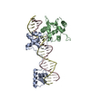
| ||||||||
|---|---|---|---|---|---|---|---|---|---|
| 1 |
| ||||||||
| Unit cell |
|
-Components
| #1: DNA chain | Mass: 8036.150 Da / Num. of mol.: 1 / Source method: obtained synthetically / Details: Synthesised by the phosphoramidite method |
|---|---|
| #2: DNA chain | Mass: 7943.119 Da / Num. of mol.: 1 / Source method: obtained synthetically / Details: Synthesised by the phosphoramidite method |
| #3: Protein | Mass: 16653.172 Da / Num. of mol.: 1 / Fragment: PAIRED DNA-BINDING DOMAIN, RESIDUES 1-149 Source method: isolated from a genetically manipulated source Source: (gene. exp.) Homo sapiens (human) / Gene: Pax5 / Plasmid: pET11a / Species (production host): Escherichia coli / Production host:  |
| #4: Protein | Mass: 18678.154 Da / Num. of mol.: 1 Fragment: INHIBITED ETS DNA-BINDING DOMAIN, RESIDUES 280-440 Source method: isolated from a genetically manipulated source Source: (gene. exp.) |
-Experimental details
-Experiment
| Experiment | Method: X-RAY DIFFRACTION / Number of used crystals: 1 |
|---|
- Sample preparation
| Crystal | Density Matthews: 2.84 Å3/Da / Density % sol: 56.73 % | ||||||||||||||||||||||||||||||||||||||||||
|---|---|---|---|---|---|---|---|---|---|---|---|---|---|---|---|---|---|---|---|---|---|---|---|---|---|---|---|---|---|---|---|---|---|---|---|---|---|---|---|---|---|---|---|
| Crystal grow | Temperature: 298 K / Method: vapor diffusion, hanging drop / pH: 5 Details: 200mM Ammonium Acetate, 10% Peg4000, 100mM Acetate buffer, pH 5.0, VAPOR DIFFUSION, HANGING DROP, temperature 298K | ||||||||||||||||||||||||||||||||||||||||||
| Crystal grow | *PLUS pH: 8 | ||||||||||||||||||||||||||||||||||||||||||
| Components of the solutions | *PLUS
|
-Data collection
| Diffraction | Mean temperature: 100 K |
|---|---|
| Diffraction source | Source: ROTATING ANODE / Type: RIGAKU RU200 / Wavelength: 1.5418 Å |
| Detector | Type: RIGAKU RAXIS IV / Detector: IMAGE PLATE / Date: Mar 13, 2001 / Details: mirrors |
| Radiation | Monochromator: Graphite / Protocol: SINGLE WAVELENGTH / Monochromatic (M) / Laue (L): M / Scattering type: x-ray |
| Radiation wavelength | Wavelength: 1.5418 Å / Relative weight: 1 |
| Reflection | Resolution: 2.8→40 Å / Num. all: 39863 / Num. obs: 13405 / % possible obs: 87.8 % / Observed criterion σ(F): 0 / Observed criterion σ(I): 2.5 / Redundancy: 3.1 % / Biso Wilson estimate: 84.6 Å2 / Rsym value: 0.06 / Net I/σ(I): 9.3 |
| Reflection shell | Resolution: 2.8→2.95 Å / Redundancy: 2.3 % / Rmerge(I) obs: 0.116 / Mean I/σ(I) obs: 4 / Num. unique all: 2937 / Rsym value: 0.116 / % possible all: 58.7 |
| Reflection | *PLUS Lowest resolution: 35 Å / Rmerge(I) obs: 0.05 |
| Reflection shell | *PLUS % possible obs: 58.7 % / Rmerge(I) obs: 0.192 |
- Processing
| Software |
| |||||||||||||||||||||||||
|---|---|---|---|---|---|---|---|---|---|---|---|---|---|---|---|---|---|---|---|---|---|---|---|---|---|---|
| Refinement | Method to determine structure: MOLECULAR REPLACEMENT Starting model: PDB ID 1K78 Resolution: 2.8→32.03 Å / Rfactor Rfree error: 0.008 / Isotropic thermal model: GROUP / Cross valid method: THROUGHOUT / σ(F): 0 / Stereochemistry target values: Engh & Huber Details: The side chains for the region 88-141 of Pax5 are included in the model despite a lack of electron density to uniquely define their location. The poor definition of the amino acid sidechains ...Details: The side chains for the region 88-141 of Pax5 are included in the model despite a lack of electron density to uniquely define their location. The poor definition of the amino acid sidechains in this region of Pax5 reflects the loss of contacts to the sugar-phosphate backbone, due to the less than optimal length of the DNA sequence used. This may explain the high B-factors obtained overall for the structure. The electron density for the side chains of residues in the region between residues 309-318 in Ets-1 was poorly defined and only the side chains for Arg309, Asp310 and Leu314 could be built.
| |||||||||||||||||||||||||
| Solvent computation | Solvent model: FLAT MODEL / Bsol: 21.8329 Å2 / ksol: 0.276781 e/Å3 | |||||||||||||||||||||||||
| Displacement parameters | Biso mean: 73.2 Å2
| |||||||||||||||||||||||||
| Refine analyze |
| |||||||||||||||||||||||||
| Refinement step | Cycle: LAST / Resolution: 2.8→32.03 Å
| |||||||||||||||||||||||||
| Refine LS restraints |
| |||||||||||||||||||||||||
| LS refinement shell | Resolution: 2.8→2.98 Å / Rfactor Rfree error: 0.034 / Total num. of bins used: 6
| |||||||||||||||||||||||||
| Xplor file |
| |||||||||||||||||||||||||
| Refinement | *PLUS Highest resolution: 2.8 Å / Lowest resolution: 35 Å / Rfactor Rfree: 0.31 | |||||||||||||||||||||||||
| Solvent computation | *PLUS | |||||||||||||||||||||||||
| Displacement parameters | *PLUS | |||||||||||||||||||||||||
| Refine LS restraints | *PLUS
|
