 Movie
Movie Controller


+ Open data

- Basic information
| Entry | Database: PDB / ID: 1md0 | ||||||
|---|---|---|---|---|---|---|---|
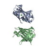
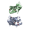
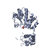
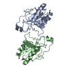
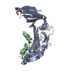
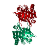
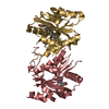
| Title | CRYSTAL STRUCTURE OF AN INHIBITED FRAGMENT OF Ets-1 | ||||||
 Components Components | C-ets-1 protein | ||||||
 Keywords Keywords | TRANSCRIPTION / AUTOINHIBITION / TRANSCRIPTION FACTOR | ||||||
| Function / homology |  Function and homology information Function and homology informationOncogene Induced Senescence / regulation of extracellular matrix disassembly / PML body organization / positive regulation of leukocyte adhesion to vascular endothelial cell / histone acetyltransferase binding / negative regulation of cell cycle / regulation of angiogenesis / positive regulation of blood vessel endothelial cell migration / immune system process / positive regulation of endothelial cell migration ...Oncogene Induced Senescence / regulation of extracellular matrix disassembly / PML body organization / positive regulation of leukocyte adhesion to vascular endothelial cell / histone acetyltransferase binding / negative regulation of cell cycle / regulation of angiogenesis / positive regulation of blood vessel endothelial cell migration / immune system process / positive regulation of endothelial cell migration / positive regulation of erythrocyte differentiation / transcription corepressor binding / cell motility / positive regulation of miRNA transcription / negative regulation of inflammatory response / positive regulation of angiogenesis / sequence-specific double-stranded DNA binding / positive regulation of inflammatory response / DNA-binding transcription activator activity, RNA polymerase II-specific / transcription regulator complex / regulation of apoptotic process / transcription by RNA polymerase II / sequence-specific DNA binding / DNA-binding transcription factor binding / nucleic acid binding / RNA polymerase II-specific DNA-binding transcription factor binding / DNA-binding transcription factor activity, RNA polymerase II-specific / cell differentiation / transcription cis-regulatory region binding / positive regulation of cell migration / RNA polymerase II cis-regulatory region sequence-specific DNA binding / DNA-binding transcription factor activity / response to antibiotic / positive regulation of cell population proliferation / positive regulation of gene expression / regulation of transcription by RNA polymerase II / positive regulation of DNA-templated transcription / positive regulation of transcription by RNA polymerase II / DNA binding / nucleoplasm / identical protein binding / nucleus / cytoplasm Similarity search - Function | ||||||
| Biological species |  | ||||||
| Method |  X-RAY DIFFRACTION / MOLECULAR REPLACEMENT / Resolution: 2 Å X-RAY DIFFRACTION / MOLECULAR REPLACEMENT / Resolution: 2 Å | ||||||
 Authors Authors | Garvie, C.W. / Pufall, M.A. / Graves, B.J. / Wolberger, C. | ||||||
 Citation Citation | Journal: J.Biol.Chem. / Year: 2002 Title: Structural Analysis of the Autoinhibition of Ets-1 and Its Role in Protein Partnerships Authors: Garvie, C.W. / Pufall, M.A. / Graves, B.J. / Wolberger, C. | ||||||
| History |
|
- Structure visualization
| Structure viewer | Molecule: MolmilJmol/JSmol |
|---|
- Downloads & links
-Download
| PDBx/mmCIF format | 1md0.cif.gz | 73.3 KB | Display | PDBx/mmCIF format |
|---|---|---|---|---|
| PDB format | pdb1md0.ent.gz | 56 KB | Display | PDB format |
| PDBx/mmJSON format | 1md0.json.gz | Tree view | PDBx/mmJSON format | |
| Others |  Other downloads Other downloads |
-Validation report
| Arichive directory | https://data.pdbj.org/pub/pdb/validation_reports/md/1md0ftp://data.pdbj.org/pub/pdb/validation_reports/md/1md0 | HTTPS FTP |
|---|
-Related structure data
| Related structure data | 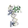 1mdmC  1k79S S: Starting model for refinement C: citing same article ( |
|---|---|
| Similar structure data |


-Links
 PDBj
PDBj- Assembly
| Deposited unit | 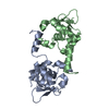
| ||||||||
|---|---|---|---|---|---|---|---|---|---|
| 1 |
| ||||||||
| Unit cell |
|
-Components
| #1: Protein | Mass: 16365.706 Da / Num. of mol.: 2 / Fragment: ETS domain, Residues 300-440 Source method: isolated from a genetically manipulated source Source: (gene. exp.)  #2: Water | ChemComp-HOH / |  Mass: 18.015 Da / Num. of mol.: 253 / Source method: isolated from a natural source / Formula: H2O Mass: 18.015 Da / Num. of mol.: 253 / Source method: isolated from a natural source / Formula: H2O |
|---|
-Experimental details
-Experiment
| Experiment | Method: X-RAY DIFFRACTION / Number of used crystals: 1 |
|---|
- Sample preparation
| Crystal | Density Matthews: 2.27 Å3/Da / Density % sol: 45.76 % | |||||||||||||||||||||||||||||||||||||||||||||||||
|---|---|---|---|---|---|---|---|---|---|---|---|---|---|---|---|---|---|---|---|---|---|---|---|---|---|---|---|---|---|---|---|---|---|---|---|---|---|---|---|---|---|---|---|---|---|---|---|---|---|---|
| Crystal grow | Temperature: 298 K / Method: vapor diffusion, hanging drop / pH: 6.5 Details: 50mM Sodium Citrate, 20% Peg4000, 100mM MES, pH 6.5, VAPOR DIFFUSION, HANGING DROP, temperature 298K | |||||||||||||||||||||||||||||||||||||||||||||||||
| Crystal grow | *PLUS pH: 8 | |||||||||||||||||||||||||||||||||||||||||||||||||
| Components of the solutions | *PLUS
|
-Data collection
| Diffraction | Mean temperature: 100 K |
|---|---|
| Diffraction source | Source: ROTATING ANODE / Type: RIGAKU RU200 / Wavelength: 1.5415 Å |
| Detector | Type: RIGAKU RAXIS IV / Detector: IMAGE PLATE / Date: Feb 1, 2001 / Details: mirrors |
| Radiation | Monochromator: graphite / Protocol: SINGLE WAVELENGTH / Monochromatic (M) / Laue (L): M / Scattering type: x-ray |
| Radiation wavelength | Wavelength: 1.5415 Å / Relative weight: 1 |
| Reflection | Resolution: 2→50 Å / Num. all: 20820 / Num. obs: 19649 / % possible obs: 94.4 % / Observed criterion σ(F): 0 / Observed criterion σ(I): -3 / Redundancy: 3.4 % / Biso Wilson estimate: 21.9 Å2 / Rmerge(I) obs: 0.055 / Rsym value: 0.055 / Net I/σ(I): 21.4 |
| Reflection shell | Resolution: 2→2.07 Å / Rmerge(I) obs: 0.176 / Mean I/σ(I) obs: 4.9 / Num. unique all: 1396 / Rsym value: 0.176 / % possible all: 68.3 |
| Reflection | *PLUS Highest resolution: 2 Å / Lowest resolution: 50 Å |
| Reflection shell | *PLUS % possible obs: 68.3 % |
- Processing
| Software |
| ||||||||||||||||||||||||||||||||||||
|---|---|---|---|---|---|---|---|---|---|---|---|---|---|---|---|---|---|---|---|---|---|---|---|---|---|---|---|---|---|---|---|---|---|---|---|---|---|
| Refinement | Method to determine structure: MOLECULAR REPLACEMENT Starting model: PDB ENTRY 1K79 Resolution: 2→41.76 Å / Rfactor Rfree error: 0.008 / Data cutoff high absF: 1491684.06 / Data cutoff high rms absF: 1491684.06 / Data cutoff low absF: 0 / Isotropic thermal model: RESTRAINED / Cross valid method: THROUGHOUT / σ(F): 0 / σ(I): 0 / Stereochemistry target values: Engh & Huber
| ||||||||||||||||||||||||||||||||||||
| Solvent computation | Solvent model: FLAT MODEL / Bsol: 46.4987 Å2 / ksol: 0.357966 e/Å3 | ||||||||||||||||||||||||||||||||||||
| Displacement parameters | Biso mean: 28.4 Å2
| ||||||||||||||||||||||||||||||||||||
| Refine analyze |
| ||||||||||||||||||||||||||||||||||||
| Refinement step | Cycle: LAST / Resolution: 2→41.76 Å
| ||||||||||||||||||||||||||||||||||||
| Refine LS restraints |
| ||||||||||||||||||||||||||||||||||||
| LS refinement shell | Resolution: 2→2.13 Å / Rfactor Rfree error: 0.029 / Total num. of bins used: 6
| ||||||||||||||||||||||||||||||||||||
| Xplor file |
| ||||||||||||||||||||||||||||||||||||
| Refinement | *PLUS Highest resolution: 2 Å / Lowest resolution: 50 Å | ||||||||||||||||||||||||||||||||||||
| Solvent computation | *PLUS | ||||||||||||||||||||||||||||||||||||
| Displacement parameters | *PLUS | ||||||||||||||||||||||||||||||||||||
| Refine LS restraints | *PLUS
|
