 Movie
Movie Controller


+ Open data

- Basic information
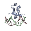
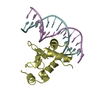
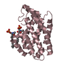
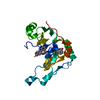
| Entry | Database: PDB / ID: 1k79 | ||||||
|---|---|---|---|---|---|---|---|
| Title | Ets-1(331-440)+GGAA duplex | ||||||
 Components Components |
| ||||||
 Keywords Keywords | TRANSCRIPTION/DNA / ETS domain / transcription factor / TRANSCRIPTION-DNA COMPLEX | ||||||
| Function / homology |  Function and homology information Function and homology informationOncogene Induced Senescence / PML body organization / regulation of extracellular matrix disassembly / positive regulation of leukocyte adhesion to vascular endothelial cell / histone acetyltransferase binding / negative regulation of cell cycle / regulation of angiogenesis / positive regulation of blood vessel endothelial cell migration / immune system process / positive regulation of erythrocyte differentiation ...Oncogene Induced Senescence / PML body organization / regulation of extracellular matrix disassembly / positive regulation of leukocyte adhesion to vascular endothelial cell / histone acetyltransferase binding / negative regulation of cell cycle / regulation of angiogenesis / positive regulation of blood vessel endothelial cell migration / immune system process / positive regulation of erythrocyte differentiation / positive regulation of endothelial cell migration / transcription corepressor binding / cell motility / negative regulation of inflammatory response / positive regulation of miRNA transcription / positive regulation of angiogenesis / positive regulation of inflammatory response / sequence-specific double-stranded DNA binding / transcription by RNA polymerase II / transcription regulator complex / DNA-binding transcription activator activity, RNA polymerase II-specific / regulation of apoptotic process / DNA-binding transcription factor binding / sequence-specific DNA binding / nucleic acid binding / RNA polymerase II-specific DNA-binding transcription factor binding / DNA-binding transcription factor activity, RNA polymerase II-specific / cell differentiation / transcription cis-regulatory region binding / positive regulation of cell migration / RNA polymerase II cis-regulatory region sequence-specific DNA binding / DNA-binding transcription factor activity / response to antibiotic / positive regulation of gene expression / positive regulation of cell population proliferation / regulation of transcription by RNA polymerase II / positive regulation of DNA-templated transcription / positive regulation of transcription by RNA polymerase II / DNA binding / nucleoplasm / identical protein binding / nucleus / cytoplasm Similarity search - Function | ||||||
| Biological species |  | ||||||
| Method |  X-RAY DIFFRACTION / MOLECULAR REPLACEMENT / Resolution: 2.4 Å X-RAY DIFFRACTION / MOLECULAR REPLACEMENT / Resolution: 2.4 Å | ||||||
 Authors Authors | Garvie, C.W. / Hagman, J. / Wolberger, C. | ||||||
 Citation Citation | Journal: Mol.Cell / Year: 2001 Title: Structural studies of Ets-1/Pax5 complex formation on DNA. Authors: Garvie, C.W. / Hagman, J. / Wolberger, C. | ||||||
| History |
|
- Structure visualization
| Structure viewer | Molecule: MolmilJmol/JSmol |
|---|
- Downloads & links
-Download
| PDBx/mmCIF format | 1k79.cif.gz | 90.1 KB | Display | PDBx/mmCIF format |
|---|---|---|---|---|
| PDB format | pdb1k79.ent.gz | 65.5 KB | Display | PDB format |
| PDBx/mmJSON format | 1k79.json.gz | Tree view | PDBx/mmJSON format | |
| Others |  Other downloads Other downloads |
-Validation report
| Arichive directory | https://data.pdbj.org/pub/pdb/validation_reports/k7/1k79ftp://data.pdbj.org/pub/pdb/validation_reports/k7/1k79 | HTTPS FTP |
|---|
-Related structure data


-Links
 PDBj
PDBj


- Assembly
| Deposited unit | 
| ||||||||
|---|---|---|---|---|---|---|---|---|---|
| 1 | 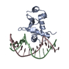
| ||||||||
| 2 | 
| ||||||||
| Unit cell |
|
-Components
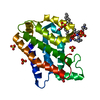
| #1: DNA chain | Mass: 4649.033 Da / Num. of mol.: 2 / Source method: obtained synthetically #2: DNA chain | Mass: 4504.936 Da / Num. of mol.: 2 / Source method: obtained synthetically #3: Protein | Mass: 12985.833 Da / Num. of mol.: 2 / Fragment: ETS domain Source method: isolated from a genetically manipulated source Source: (gene. exp.)  #4: Water | ChemComp-HOH / |  Mass: 18.015 Da / Num. of mol.: 96 / Source method: isolated from a natural source / Formula: H2O Mass: 18.015 Da / Num. of mol.: 96 / Source method: isolated from a natural source / Formula: H2O |
|---|
-Experimental details
-Experiment
| Experiment | Method: X-RAY DIFFRACTION / Number of used crystals: 1 |
|---|
- Sample preparation
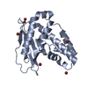
| Crystal | Density Matthews: 2.76 Å3/Da / Density % sol: 55.43 % | ||||||||||||||||||||||||
|---|---|---|---|---|---|---|---|---|---|---|---|---|---|---|---|---|---|---|---|---|---|---|---|---|---|
| Crystal grow | Temperature: 293 K / Method: vapor diffusion, hanging drop / pH: 6.5 Details: 1.4M-1.6M Sodium Citrate, 100mM Hepes, pH 6.5, VAPOR DIFFUSION, HANGING DROP, temperature 293K | ||||||||||||||||||||||||
| Components of the solutions |
| ||||||||||||||||||||||||
| Crystal grow | *PLUS | ||||||||||||||||||||||||
| Components of the solutions | *PLUS
|
-Data collection
| Diffraction | Mean temperature: 100 K |
|---|---|
| Diffraction source | Source: ROTATING ANODE / Type: RIGAKU RU200 / Wavelength: 1.5418 Å |
| Detector | Type: RIGAKU RAXIS IV / Detector: IMAGE PLATE / Date: Feb 6, 2001 / Details: mirrors |
| Radiation | Monochromator: Graphite / Protocol: SINGLE WAVELENGTH / Monochromatic (M) / Laue (L): M / Scattering type: x-ray |
| Radiation wavelength | Wavelength: 1.5418 Å / Relative weight: 1 |
| Reflection | Resolution: 2.4→17.05 Å / Num. all: 17326 / Num. obs: 17326 / % possible obs: 89.9 % / Observed criterion σ(F): 0 / Observed criterion σ(I): 0 / Redundancy: 3.4 % / Biso Wilson estimate: 47.9 Å2 / Rmerge(I) obs: 0.061 / Net I/σ(I): 18.7 |
| Reflection shell | Resolution: 2.4→2.49 Å / Rmerge(I) obs: 0.278 / Mean I/σ(I) obs: 2.7 / % possible all: 58.7 |
| Reflection | *PLUS |
| Reflection shell | *PLUS % possible obs: 58.7 % |
- Processing
| Software |
| ||||||||||||||||||||||||||||||||||||||||
|---|---|---|---|---|---|---|---|---|---|---|---|---|---|---|---|---|---|---|---|---|---|---|---|---|---|---|---|---|---|---|---|---|---|---|---|---|---|---|---|---|---|
| Refinement | Method to determine structure: MOLECULAR REPLACEMENT Starting model: Ets-1(331-440) bound to DNA from the Pax5(1-149)+Ets-1(331-440)+DNA complex Resolution: 2.4→17.05 Å / Rfactor Rfree error: 0.007 / Data cutoff high absF: 1024028 / Data cutoff low absF: 0 / Isotropic thermal model: RESTRAINED / Cross valid method: THROUGHOUT / σ(F): 0 / σ(I): 0 / Stereochemistry target values: ENGH AND HUBER
| ||||||||||||||||||||||||||||||||||||||||
| Solvent computation | Solvent model: FLAT MODEL / Bsol: 42.0221 Å2 / ksol: 0.370139 e/Å3 | ||||||||||||||||||||||||||||||||||||||||
| Displacement parameters | Biso mean: 44.6 Å2
| ||||||||||||||||||||||||||||||||||||||||
| Refine analyze |
| ||||||||||||||||||||||||||||||||||||||||
| Refinement step | Cycle: LAST / Resolution: 2.4→17.05 Å
| ||||||||||||||||||||||||||||||||||||||||
| Refine LS restraints |
| ||||||||||||||||||||||||||||||||||||||||
| LS refinement shell | Resolution: 2.4→2.55 Å / Rfactor Rfree error: 0.029 / Total num. of bins used: 6
| ||||||||||||||||||||||||||||||||||||||||
| Xplor file |
| ||||||||||||||||||||||||||||||||||||||||
| Software | *PLUS Name: CNS / Version: 1 / Classification: refinement | ||||||||||||||||||||||||||||||||||||||||
| Refinement | *PLUS Lowest resolution: 17.1 Å / σ(F): 0 / % reflection Rfree: 10 % / Rfactor obs: 0.228 / Rfactor Rfree: 0.275 | ||||||||||||||||||||||||||||||||||||||||
| Solvent computation | *PLUS | ||||||||||||||||||||||||||||||||||||||||
| Displacement parameters | *PLUS Biso mean: 44.6 Å2 | ||||||||||||||||||||||||||||||||||||||||
| Refine LS restraints | *PLUS
| ||||||||||||||||||||||||||||||||||||||||
| LS refinement shell | *PLUS Rfactor Rfree: 0.419 / Rfactor Rwork: 0.373 |
