 Movie
Movie Controller

[English] 日本語
 Yorodumi
- PDB-6kny: Structure of Amuc_1100 without transmembrane region from Akkerman... -
+ Open data

- Basic information
| Entry | Database: PDB / ID: 6kny | |||||||||
|---|---|---|---|---|---|---|---|---|---|---|
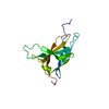
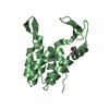
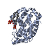
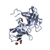
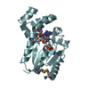
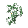
| Title | Structure of Amuc_1100 without transmembrane region from Akkermansia muciniphila | |||||||||
 Components Components | Protein Amuc_1100 | |||||||||
 Keywords Keywords | UNKNOWN FUNCTION / Uncharacterized / protein | |||||||||
| Function / homology | : / Amuc_1100 pilus-like / Uncharacterized protein Function and homology information Function and homology information | |||||||||
| Biological species |  Akkermansia muciniphila (bacteria) Akkermansia muciniphila (bacteria) | |||||||||
| Method |  X-RAY DIFFRACTION / SYNCHROTRON / SAD / Resolution: 2.1 Å X-RAY DIFFRACTION / SYNCHROTRON / SAD / Resolution: 2.1 Å | |||||||||
 Authors Authors | Mou, L.Q. / Xiao, Q.J. / Deng, D. | |||||||||
| Funding support |  China, 2items China, 2items
| |||||||||
 Citation Citation | Journal: Acta Crystallogr.,Sect.F / Year: 2020 Title: Crystal structure of monomeric Amuc_1100 from Akkermansia muciniphila. Authors: Mou, L. / Peng, X. / Chen, Y. / Xiao, Q. / Liao, H. / Liu, M. / Guo, L. / Liu, Y. / Zhang, X. / Deng, D. | |||||||||
| History |
|
- Structure visualization
| Structure viewer | Molecule: MolmilJmol/JSmol |
|---|
- Downloads & links
-Download
| PDBx/mmCIF format | 6kny.cif.gz | 129 KB | Display | PDBx/mmCIF format |
|---|---|---|---|---|
| PDB format | pdb6kny.ent.gz | 93.8 KB | Display | PDB format |
| PDBx/mmJSON format | 6kny.json.gz | Tree view | PDBx/mmJSON format | |
| Others |  Other downloads Other downloads |
-Validation report
| Arichive directory | https://data.pdbj.org/pub/pdb/validation_reports/kn/6knyftp://data.pdbj.org/pub/pdb/validation_reports/kn/6kny | HTTPS FTP |
|---|
-Related structure data
| Similar structure data |
|---|


-Links
 PDBj
PDBj- Assembly
| Deposited unit | 
| ||||||||||||
|---|---|---|---|---|---|---|---|---|---|---|---|---|---|
| 1 | 
| ||||||||||||
| 2 | 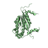
| ||||||||||||
| 3 | 
| ||||||||||||
| 4 |
| ||||||||||||
| Unit cell |
| ||||||||||||
| Components on special symmetry positions |
|
-Components
| #1: Protein | Mass: 31150.359 Da / Num. of mol.: 2 Source method: isolated from a genetically manipulated source Source: (gene. exp.) Akkermansia muciniphila (strain ATCC BAA-835 / Muc) (bacteria)Gene: Amuc_1100 / Production host: #2: Water | ChemComp-HOH / |  Mass: 18.015 Da / Num. of mol.: 169 / Source method: isolated from a natural source / Formula: H2O Mass: 18.015 Da / Num. of mol.: 169 / Source method: isolated from a natural source / Formula: H2O |
|---|
-Experimental details
-Experiment
| Experiment | Method: X-RAY DIFFRACTION / Number of used crystals: 1 |
|---|
- Sample preparation
| Crystal | Density Matthews: 1.81 Å3/Da / Density % sol: 32.23 % |
|---|---|
| Crystal grow | Temperature: 297 K / Method: vapor diffusion, sitting drop Details: 0.2M potassium phosphate dibasic, 1.8 M sodium phosphate monobasic, 0.1M sodium phosphate dibasic/citric acid pH 4.2 |
-Data collection
| Diffraction | Mean temperature: 100 K / Serial crystal experiment: N | |||||||||||||||||||||||||||||||||||||||||||||||||||||||||||||||||||||||||||||||||||||||||||||||||||
|---|---|---|---|---|---|---|---|---|---|---|---|---|---|---|---|---|---|---|---|---|---|---|---|---|---|---|---|---|---|---|---|---|---|---|---|---|---|---|---|---|---|---|---|---|---|---|---|---|---|---|---|---|---|---|---|---|---|---|---|---|---|---|---|---|---|---|---|---|---|---|---|---|---|---|---|---|---|---|---|---|---|---|---|---|---|---|---|---|---|---|---|---|---|---|---|---|---|---|---|---|
| Diffraction source | Source: SYNCHROTRON / Site: SSRF / Beamline: BL18U1 / Wavelength: 0.9791 Å | |||||||||||||||||||||||||||||||||||||||||||||||||||||||||||||||||||||||||||||||||||||||||||||||||||
| Detector | Type: DECTRIS PILATUS 6M / Detector: PIXEL / Date: Nov 20, 2018 | |||||||||||||||||||||||||||||||||||||||||||||||||||||||||||||||||||||||||||||||||||||||||||||||||||
| Radiation | Protocol: SINGLE WAVELENGTH / Monochromatic (M) / Laue (L): M / Scattering type: x-ray | |||||||||||||||||||||||||||||||||||||||||||||||||||||||||||||||||||||||||||||||||||||||||||||||||||
| Radiation wavelength | Wavelength: 0.9791 Å / Relative weight: 1 | |||||||||||||||||||||||||||||||||||||||||||||||||||||||||||||||||||||||||||||||||||||||||||||||||||
| Reflection | Resolution: 2.094→40 Å / Num. obs: 26899 / % possible obs: 100 % / Redundancy: 6.4 % / Biso Wilson estimate: 30.99 Å2 / Rmerge(I) obs: 0.137 / Rpim(I) all: 0.058 / Rrim(I) all: 0.149 / Χ2: 0.546 / Net I/σ(I): 3.1 | |||||||||||||||||||||||||||||||||||||||||||||||||||||||||||||||||||||||||||||||||||||||||||||||||||
| Reflection shell | Diffraction-ID: 1
|
-Phasing
| Phasing | Method: SAD |
|---|
- Processing
| Software |
| ||||||||||||||||||||||||||||||||||||||||||||||||||||||||||||||||||||||||||||||||||||||||||
|---|---|---|---|---|---|---|---|---|---|---|---|---|---|---|---|---|---|---|---|---|---|---|---|---|---|---|---|---|---|---|---|---|---|---|---|---|---|---|---|---|---|---|---|---|---|---|---|---|---|---|---|---|---|---|---|---|---|---|---|---|---|---|---|---|---|---|---|---|---|---|---|---|---|---|---|---|---|---|---|---|---|---|---|---|---|---|---|---|---|---|---|
| Refinement | Method to determine structure: SAD / Resolution: 2.1→38.991 Å / SU ML: 0.15 / Cross valid method: THROUGHOUT / σ(F): 1.37 / Phase error: 18.88
| ||||||||||||||||||||||||||||||||||||||||||||||||||||||||||||||||||||||||||||||||||||||||||
| Solvent computation | Shrinkage radii: 0.9 Å / VDW probe radii: 1.11 Å | ||||||||||||||||||||||||||||||||||||||||||||||||||||||||||||||||||||||||||||||||||||||||||
| Displacement parameters | Biso max: 114.34 Å2 / Biso mean: 34.8405 Å2 / Biso min: 19.32 Å2 | ||||||||||||||||||||||||||||||||||||||||||||||||||||||||||||||||||||||||||||||||||||||||||
| Refinement step | Cycle: final / Resolution: 2.1→38.991 Å
| ||||||||||||||||||||||||||||||||||||||||||||||||||||||||||||||||||||||||||||||||||||||||||
| LS refinement shell | Refine-ID: X-RAY DIFFRACTION / Rfactor Rfree error: 0
|
