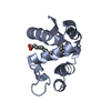
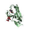
Entry Database : PDB / ID : 1m9zTitle CRYSTAL STRUCTURE OF HUMAN TGF-BETA TYPE II RECEPTOR LIGAND BINDING DOMAIN TGF-BETA RECEPTOR TYPE II Keywords / / Function / homology Function Domain/homology Component
/ / / / / / / / / / / / / / / / / / / / / / / / / / / / / / / / / / / / / / / / / / / / / / / / / / / / / / / / / / / / / / / / / / / / / / / / / / / / / / / / / / / / / / / / / / / / / / / / / / / / / / / / / / / / / / / / / / / / / / / / / / / / / / / / / / / / / / / Biological species Homo sapiens (human)Method / / / Resolution : 1.05 Å Authors Boesen, C.C. / Radaev, S. / Motyka, S.A. / Patamawenu, A. / Sun, P.D. Journal : Structure / Year : 2002Title : THE 1.1A CRYSTAL STRUCTURE OF HUMAN TGF-BETA TYPE II RECEPTOR LIGAND BINDING DOMAINAuthors : Boesen, C.C. / Radaev, S. / Motyka, S.A. / Patamawenu, A. / Sun, P.D. History Deposition Jul 30, 2002 Deposition site / Processing site Revision 1.0 Sep 11, 2002 Provider / Type Revision 1.1 Apr 28, 2008 Group Revision 1.2 Jul 13, 2011 Group / Version format complianceRevision 1.3 Jul 24, 2019 Group / Data collection / Refinement description / Category / softwareItem / _software.name / _software.versionRevision 1.4 Oct 27, 2021 Group / Database references / Derived calculationsCategory database_2 / pdbx_unobs_or_zero_occ_atoms ... database_2 / pdbx_unobs_or_zero_occ_atoms / struct_ref_seq_dif / struct_site Item _database_2.pdbx_DOI / _database_2.pdbx_database_accession ... _database_2.pdbx_DOI / _database_2.pdbx_database_accession / _struct_ref_seq_dif.details / _struct_site.pdbx_auth_asym_id / _struct_site.pdbx_auth_comp_id / _struct_site.pdbx_auth_seq_id Revision 1.5 Oct 16, 2024 Group / Structure summaryCategory chem_comp_atom / chem_comp_bond ... chem_comp_atom / chem_comp_bond / pdbx_entry_details / pdbx_modification_feature
Show all Show less
 Movie
Movie Controller

 Yorodumi
 Open data
 Basic information
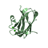
 Components
 Keywords
 Function and homology information
 Homo sapiens (human)
 X-RAY DIFFRACTION /
  Authors
 Citation
 Structure visualization
 Downloads & links
 Other downloads


 PDBj
PDBj


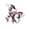
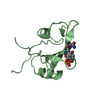
 Assembly
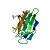


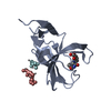
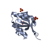
 Mass: 92.094 Da / Num. of mol.: 1 / Source method: obtained synthetically / Formula: C3H8O3
 Mass: 18.015 Da / Num. of mol.: 178 / Source method: isolated from a natural source / Formula: H2O
 Sample preparation
 / Beamline: X9B / Wavelength: 1.0092, 1.0011, 1.0076
 Processing