- PDB-1m6i: Crystal Structure of Apoptosis Inducing Factor (AIF) -
+
Open data
ID or keywords:
Loading...
-
Basic information
Entry
Database: PDB / ID: 1m6i
Title
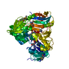
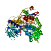
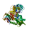
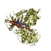
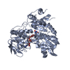
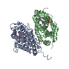
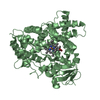
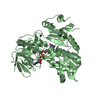
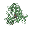
Crystal Structure of Apoptosis Inducing Factor (AIF)
Components
Programmed cell death protein 8
Keywords
OXIDOREDUCTASE / Apoptosis / AIF
Function / homology
Function and homology information
Oxidoreductases; Acting on NADH or NADPH; With unknown physiological acceptors / protein import into the intermembrane space via the disulfide relay system / cellular response to aldosterone / mitochondrial respiratory chain complex assembly / protein import into mitochondrial intermembrane space / poly-ADP-D-ribose binding / NAD(P)H oxidase H2O2-forming activity / positive regulation of necroptotic process / response to L-glutamate / intrinsic apoptotic signaling pathway in response to endoplasmic reticulum stress ...Oxidoreductases; Acting on NADH or NADPH; With unknown physiological acceptors / protein import into the intermembrane space via the disulfide relay system / cellular response to aldosterone / mitochondrial respiratory chain complex assembly / protein import into mitochondrial intermembrane space / poly-ADP-D-ribose binding / NAD(P)H oxidase H2O2-forming activity / positive regulation of necroptotic process / response to L-glutamate / intrinsic apoptotic signaling pathway in response to endoplasmic reticulum stress / oxidoreductase activity, acting on NAD(P)H / NADH dehydrogenase activity / sperm head-tail coupling apparatus / cellular response to nitric oxide / FAD binding / response to ischemia / cellular response to estradiol stimulus / mitochondrial intermembrane space / response to toxic substance / cellular response to hydrogen peroxide / neuron differentiation / positive regulation of neuron apoptotic process / cellular response to hypoxia / protein dimerization activity / mitochondrial inner membrane / positive regulation of apoptotic process / apoptotic process / perinuclear region of cytoplasm / mitochondrion / DNA binding / nucleus / cytosol Similarity search - Function
In the structure databanks used in Yorodumi, some data are registered as the other names, "COVID-19 virus" and "2019-nCoV". Here are the details of the virus and the list of structure data.
Jan 31, 2019. EMDB accession codes are about to change! (news from PDBe EMDB page)
EMDB accession codes are about to change! (news from PDBe EMDB page)
The allocation of 4 digits for EMDB accession codes will soon come to an end. Whilst these codes will remain in use, new EMDB accession codes will include an additional digit and will expand incrementally as the available range of codes is exhausted. The current 4-digit format prefixed with “EMD-” (i.e. EMD-XXXX) will advance to a 5-digit format (i.e. EMD-XXXXX), and so on. It is currently estimated that the 4-digit codes will be depleted around Spring 2019, at which point the 5-digit format will come into force.
The EM Navigator/Yorodumi systems omit the EMD- prefix.
Related info.:Q: What is EMD? / ID/Accession-code notation in Yorodumi/EM Navigator
Yorodumi is a browser for structure data from EMDB, PDB, SASBDB, etc.
This page is also the successor to EM Navigator detail page, and also detail information page/front-end page for Omokage search.
The word "yorodu" (or yorozu) is an old Japanese word meaning "ten thousand". "mi" (miru) is to see.
Related info.:EMDB / PDB / SASBDB / Comparison of 3 databanks / Yorodumi Search / Aug 31, 2016. New EM Navigator & Yorodumi / Yorodumi Papers / Jmol/JSmol / Function and homology information / Changes in new EM Navigator and Yorodumi
 Movie
Movie Controller


 Open data
 Basic information
 Components
 Keywords
 Function and homology information
 Homo sapiens (human)
 X-RAY DIFFRACTION /
  Authors
 Citation
 Structure visualization
 Downloads & links
 Other downloads

 PDBj
PDBj


 Assembly
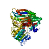


 Mass: 785.550 Da / Num. of mol.: 1 / Source method: obtained synthetically / Formula: C27H33N9O15P2 / Comment: FAD*YM
 Mass: 18.015 Da / Num. of mol.: 235 / Source method: isolated from a natural source / Formula: H2O
 Sample preparation
 / Beamline: 19-ID / Wavelength: 0.9997 Å
 Processing