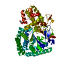
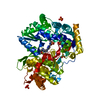
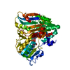
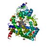
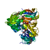
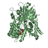
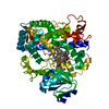
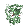
Entry Database : PDB / ID : 3uvvTitle Crystal Structure of the ligand binding domains of the thyroid receptor:retinoid X receptor complexed with 3,3',5 triiodo-L-thyronine and 9-cis retinoic acid Retinoic acid receptor RXR-alpha Thyroid hormone receptor alpha Keywords / / / / / Function / homology Function Domain/homology Component
/ / / / / / / / / / / / / / / / / / / / / / / / / / / / / / / / / / / / / / / / / / / / / / / / / / / / / / / / / / / / / / / / / / / / / / / / / / / / / / / / / / / / / / / / / / / / / / / / / / / / / / / / / / / / / / / / / / / / / / / / / / Biological species Gallus gallus (chicken)Homo sapiens (human)Method / / / Resolution : 2.95 Å Authors Fernandez, E.J. / Putcha, B.-D.K. / Wright, E. / Brunzelle, J.S. Journal : Proc.Natl.Acad.Sci.USA / Year : 2012Title : Structural basis for negative cooperativity within agonist-bound TR:RXR heterodimers.Authors : Putcha, B.D. / Wright, E. / Brunzelle, J.S. / Fernandez, E.J. History Deposition Nov 30, 2011 Deposition site / Processing site Revision 1.0 Apr 18, 2012 Provider / Type Revision 1.1 May 2, 2012 Group Revision 1.2 Nov 16, 2016 Group Revision 1.3 Sep 13, 2023 Group Data collection / Database references ... Data collection / Database references / Derived calculations / Refinement description Category chem_comp_atom / chem_comp_bond ... chem_comp_atom / chem_comp_bond / database_2 / pdbx_initial_refinement_model / struct_ref_seq_dif / struct_site Item _database_2.pdbx_DOI / _database_2.pdbx_database_accession ... _database_2.pdbx_DOI / _database_2.pdbx_database_accession / _struct_ref_seq_dif.details / _struct_site.pdbx_auth_asym_id / _struct_site.pdbx_auth_comp_id / _struct_site.pdbx_auth_seq_id Revision 2.0 Nov 15, 2023 Group / Data collection / Category / chem_comp_atom / chem_comp_bondItem _atom_site.auth_atom_id / _atom_site.label_atom_id ... _atom_site.auth_atom_id / _atom_site.label_atom_id / _chem_comp_atom.atom_id / _chem_comp_bond.atom_id_1 / _chem_comp_bond.atom_id_2
Show all Show less
 Movie
Movie Controller

 Yorodumi
 Open data
 Basic information
 Components
 Keywords
 Function and homology information
 Homo sapiens (human)
 X-RAY DIFFRACTION /
  Authors
 Citation
 Structure visualization
 Downloads & links
 Other downloads
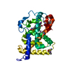


 PDBj
PDBj


 Assembly
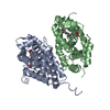

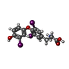
 Mass: 650.973 Da / Num. of mol.: 1 / Source method: obtained synthetically / Formula: C15H12I3NO4 / Comment: hormone*YM
 Mass: 300.435 Da / Num. of mol.: 1 / Source method: obtained synthetically / Formula: C20H28O2
 Mass: 18.015 Da / Num. of mol.: 19 / Source method: isolated from a natural source / Formula: H2O
 Sample preparation
 / Beamline: 21-ID-D / Wavelength: 1.54984 Å
 Processing