 Movie
Movie Controller


+ Open data

- Basic information
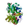
| Entry | Database: PDB / ID: 1m54 | ||||||
|---|---|---|---|---|---|---|---|
| Title | CYSTATHIONINE-BETA SYNTHASE: REDUCED VICINAL THIOLS | ||||||
 Components Components | CYSTATHIONINE BETA-SYNTHASE | ||||||
 Keywords Keywords | LYASE / PLP PROTEIN FOLD TYPE II (TRYPTOPHAN SYNTHASE) / PLP AND HEME BOUND TO PROTEIN / REDUCED VICINAL CYSTEINES | ||||||
| Function / homology |  Function and homology information Function and homology informationCysteine formation from homocysteine / L-homocysteine catabolic process / cerebellum morphogenesis / modified amino acid binding / cystathionine beta-synthase / cystathionine beta-synthase activity / L-serine catabolic process / : / Metabolism of ingested SeMet, Sec, MeSec into H2Se / : ...Cysteine formation from homocysteine / L-homocysteine catabolic process / cerebellum morphogenesis / modified amino acid binding / cystathionine beta-synthase / cystathionine beta-synthase activity / L-serine catabolic process / : / Metabolism of ingested SeMet, Sec, MeSec into H2Se / : / carbon monoxide binding / cartilage development involved in endochondral bone morphogenesis / hydrogen sulfide biosynthetic process / homocysteine metabolic process / L-cysteine catabolic process / L-serine metabolic process / L-cysteine biosynthetic process / endochondral ossification / response to folic acid / transsulfuration / nitric oxide binding / : / DNA protection / S-adenosyl-L-methionine binding / nitrite reductase (NO-forming) activity / regulation of JNK cascade / superoxide metabolic process / maternal process involved in female pregnancy / blood vessel remodeling / blood vessel diameter maintenance / oxygen binding / pyridoxal phosphate binding / cellular response to hypoxia / heme binding / ubiquitin protein ligase binding / negative regulation of apoptotic process / enzyme binding / protein homodimerization activity / metal ion binding / identical protein binding / nucleus / cytosol / cytoplasm Similarity search - Function | ||||||
| Biological species |  Homo sapiens (human) Homo sapiens (human) | ||||||
| Method |  X-RAY DIFFRACTION / SYNCHROTRON / MOLECULAR REPLACEMENT / Resolution: 2.9 Å X-RAY DIFFRACTION / SYNCHROTRON / MOLECULAR REPLACEMENT / Resolution: 2.9 Å | ||||||
 Authors Authors | Taoka, S. / Lepore, B.W. / Kabil, O. / Ojha, S. / Ringe, D. / Banerjee, R. | ||||||
 Citation Citation | Journal: BIOCHEMISTRY / Year: 2002 Title: HUMAN CYSTATHIONINE BETA-SYNTHASE IS A HEME SENSOR PROTEIN. EVIDENCE THAT THE REDOX SENSOR IS HEME AND NOT THE VICINAL CYSTEINES IN THE CXXC MOTIF SEEN IN THE CRYSTAL STRUCTURE OF THE TRUNCATED ENZYME Authors: Taoka, S. / Lepore, B.W. / Kabil, O. / Ojha, S. / Ringe, D. / Banerjee, R. #1: Journal: Embo J. / Year: 2001Title: STRUCTURE OF HUMAN CYSTATHIONINE-SYNTHASE: A UNIQUE PYRIDOXAL 5'-PHOSPHATE-DEPENDENT HEME PROTEIN Authors: Meier, M. / Janosik, M. / Kery, V. / Kraus, J.P. / Burkhard, P. | ||||||
| History |
|
- Structure visualization
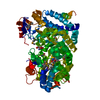
| Structure viewer | Molecule: MolmilJmol/JSmol |
|---|
- Downloads & links
-Download
| PDBx/mmCIF format | 1m54.cif.gz | 407.5 KB | Display | PDBx/mmCIF format |
|---|---|---|---|---|
| PDB format | pdb1m54.ent.gz | 332.4 KB | Display | PDB format |
| PDBx/mmJSON format | 1m54.json.gz | Tree view | PDBx/mmJSON format | |
| Others |  Other downloads Other downloads |
-Validation report
| Arichive directory | https://data.pdbj.org/pub/pdb/validation_reports/m5/1m54ftp://data.pdbj.org/pub/pdb/validation_reports/m5/1m54 | HTTPS FTP |
|---|
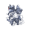
-Related structure data
| Related structure data | 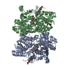 1jbqS S: Starting model for refinement |
|---|---|
| Similar structure data |


-Links
 PDBj
PDBj

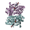
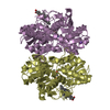
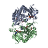
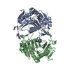
- Assembly
| Deposited unit | 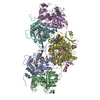
| ||||||||
|---|---|---|---|---|---|---|---|---|---|
| 1 | 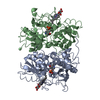
| ||||||||
| 2 | 
| ||||||||
| 3 | 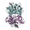
| ||||||||
| Unit cell |
| ||||||||
| Details | the biologically relevant entity is a dimer. three dimers occupy the assymetric unit. chain A is related to chain B by the followin 3x3 matrix: ( 0.78094 -0.51950 -0.34677 ) ( -0.51627 -0.84936 0.10976 ) ( -0.35155 0.09331 -0.93151 ) translation: (-0.12746 -0.20056 -0.68113) |
-Components
| #1: Protein | Mass: 39922.727 Da / Num. of mol.: 6 / Fragment: RESIDUES 44-406 / Mutation: I44M Source method: isolated from a genetically manipulated source Source: (gene. exp.) Homo sapiens (human) / Gene: CBS / Organ: blood / Plasmid: pCBS-deltaN43/deltaC143 / Species (production host): Escherichia coli / Production host:  #2: Chemical | ChemComp-PLP /   Mass: 247.142 Da / Num. of mol.: 6 / Source method: obtained synthetically / Formula: C8H10NO6P Mass: 247.142 Da / Num. of mol.: 6 / Source method: obtained synthetically / Formula: C8H10NO6P#3: Chemical | ChemComp-HEM /   Mass: 616.487 Da / Num. of mol.: 6 / Source method: obtained synthetically / Formula: C34H32FeN4O4 Mass: 616.487 Da / Num. of mol.: 6 / Source method: obtained synthetically / Formula: C34H32FeN4O4#4: Water | ChemComp-HOH / |  Mass: 18.015 Da / Num. of mol.: 104 / Source method: isolated from a natural source / Formula: H2O Mass: 18.015 Da / Num. of mol.: 104 / Source method: isolated from a natural source / Formula: H2OHas protein modification | N | |
|---|
-Experimental details
-Experiment
| Experiment | Method: X-RAY DIFFRACTION / Number of used crystals: 1 |
|---|
- Sample preparation
| Crystal | Density Matthews: 2.55 Å3/Da / Density % sol: 51.83 % | ||||||||||||||||||||||||||||||||||||
|---|---|---|---|---|---|---|---|---|---|---|---|---|---|---|---|---|---|---|---|---|---|---|---|---|---|---|---|---|---|---|---|---|---|---|---|---|---|
| Crystal grow | Temperature: 323 K / Method: vapor diffusion, hanging drop / pH: 8 Details: PEG 10K, Tris-HCl, pH 8.0, VAPOR DIFFUSION, HANGING DROP, temperature 323K | ||||||||||||||||||||||||||||||||||||
| Crystal grow | *PLUS Temperature: 25 ℃ | ||||||||||||||||||||||||||||||||||||
| Components of the solutions | *PLUS
|
-Data collection
| Diffraction | Mean temperature: 100 K | |||||||||
|---|---|---|---|---|---|---|---|---|---|---|
| Diffraction source | Source: SYNCHROTRON / Site: APS  / Beamline: 14-BM-D / Wavelength: 1.7401, 1.7372 / Beamline: 14-BM-D / Wavelength: 1.7401, 1.7372 | |||||||||
| Detector | Type: ADSC QUANTUM 4 / Detector: CCD / Date: Mar 16, 2001 / Details: Bent cylindrical Si-mirror (Rh coating) | |||||||||
| Radiation | Monochromator: Si(111) double-crystal monochromator / Protocol: MAD / Monochromatic (M) / Laue (L): M / Scattering type: x-ray | |||||||||
| Radiation wavelength |
| |||||||||
| Reflection | Resolution: 2.9→50 Å / Num. all: 87507 / Num. obs: 87507 / % possible obs: 83.2 % / Observed criterion σ(I): -3 / Redundancy: 12 % / Biso Wilson estimate: 58.127 Å2 / Rmerge(I) obs: 0.14 / Rsym value: 0.14 / Net I/σ(I): 9.2 | |||||||||
| Reflection shell | Resolution: 2.9→3 Å / Redundancy: 10 % / Rmerge(I) obs: 0.459 / Mean I/σ(I) obs: 3 / Num. unique all: 53028 / Rsym value: 0.459 / % possible all: 10 | |||||||||
| Reflection | *PLUS Highest resolution: 2.9 Å / Num. obs: 53028 / % possible obs: 93.8 % / Num. measured all: 628994 / Rmerge(I) obs: 0.14 | |||||||||
| Reflection shell | *PLUS Highest resolution: 2.9 Å / % possible obs: 93.2 % / Rmerge(I) obs: 0.459 |
- Processing
| Software |
| |||||||||||||||||||||||||
|---|---|---|---|---|---|---|---|---|---|---|---|---|---|---|---|---|---|---|---|---|---|---|---|---|---|---|
| Refinement | Method to determine structure: MOLECULAR REPLACEMENT Starting model: PDB ENTRY 1JBQ Resolution: 2.9→50 Å / Isotropic thermal model: isotropic / Cross valid method: THROUGHOUT / σ(F): 1 / σ(I): 1 / Stereochemistry target values: Engh & Huber
| |||||||||||||||||||||||||
| Displacement parameters | Biso mean: 33.6 Å2
| |||||||||||||||||||||||||
| Refine analyze |
| |||||||||||||||||||||||||
| Refinement step | Cycle: LAST / Resolution: 2.9→50 Å
| |||||||||||||||||||||||||
| Refine LS restraints |
| |||||||||||||||||||||||||
| LS refinement shell | Resolution: 2.9→3.08 Å / Rfactor Rfree error: 0.012
| |||||||||||||||||||||||||
| Refinement | *PLUS Highest resolution: 2.9 Å / Lowest resolution: 50 Å / Num. reflection all: 628994 / Num. reflection obs: 53028 / Num. reflection Rfree: 8143 / % reflection Rfree: 7.7 % / Rfactor Rfree: 0.348 / Rfactor Rwork: 0.259 | |||||||||||||||||||||||||
| Solvent computation | *PLUS | |||||||||||||||||||||||||
| Displacement parameters | *PLUS | |||||||||||||||||||||||||
| Refine LS restraints | *PLUS
| |||||||||||||||||||||||||
| LS refinement shell | *PLUS Rfactor Rfree: 0.405 / Rfactor Rwork: 0.336 |
