 Movie
Movie Controller

[English] 日本語
 Yorodumi
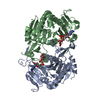
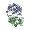
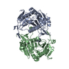
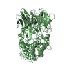
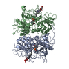
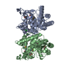
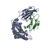
- PDB-1z84: X-ray structure of galt-like protein from arabidopsis thaliana at... -
+ Open data

- Basic information
| Entry | Database: PDB / ID: 1z84 | ||||||
|---|---|---|---|---|---|---|---|
| Title | X-ray structure of galt-like protein from arabidopsis thaliana at5g18200 | ||||||
 Components Components | galactose-1-phosphate uridyl transferase-like protein | ||||||
 Keywords Keywords | STRUCTURAL GENOMICS / UNKNOWN FUNCTION / GALT / ZINC / AMP / PROTEIN STRUCTURE INITIATIVE / PSI / CESG / CENTER FOR EUKARYOTIC STRUCTURAL GENOMICS | ||||||
| Function / homology |  Function and homology information Function and homology informationribose-5-phosphate adenylyltransferase activity / positive regulation of cellular response to phosphate starvation / UDP-glucose:hexose-1-phosphate uridylyltransferase activity / galactose metabolic process / nucleotidyltransferase activity / glucose metabolic process / ADP binding / Transferases; Transferring phosphorus-containing groups; Nucleotidyltransferases / carbohydrate metabolic process / zinc ion binding Similarity search - Function | ||||||
| Biological species |  | ||||||
| Method |  X-RAY DIFFRACTION / SYNCHROTRON / MOLECULAR REPLACEMENT / molecular replacement / Resolution: 1.83 Å X-RAY DIFFRACTION / SYNCHROTRON / MOLECULAR REPLACEMENT / molecular replacement / Resolution: 1.83 Å | ||||||
 Authors Authors | Mccoy, J.G. / Bitto, E. / Phillips Jr., G.N. / Bingman, C.A. / Center for Eukaryotic Structural Genomics (CESG) | ||||||
 Citation Citation | Journal: Biochemistry / Year: 2006 Title: Structure and Mechanism of an ADP-Glucose Phosphorylase from Arabidopsis thaliana Authors: MCCOY, J.G. / ARABSHAHI, A. / BITTO, E. / BINGMAN, C.A. / RUZICKA, F.J. / FREY, P.A. / PHILLIPS JR., G.N. | ||||||
| History |
|
- Structure visualization
| Structure viewer | Molecule: MolmilJmol/JSmol |
|---|
- Downloads & links
-Download
| PDBx/mmCIF format | 1z84.cif.gz | 149.6 KB | Display | PDBx/mmCIF format |
|---|---|---|---|---|
| PDB format | pdb1z84.ent.gz | 113.6 KB | Display | PDB format |
| PDBx/mmJSON format | 1z84.json.gz | Tree view | PDBx/mmJSON format | |
| Others |  Other downloads Other downloads |
-Validation report
| Arichive directory | https://data.pdbj.org/pub/pdb/validation_reports/z8/1z84ftp://data.pdbj.org/pub/pdb/validation_reports/z8/1z84 | HTTPS FTP |
|---|
-Related structure data
| Related structure data |  1zwjC  1vkv C: citing same article ( S: Starting model for refinement |
|---|---|
| Similar structure data | |
| Other databases |


-Links
 PDBj
PDBj


- Assembly
| Deposited unit | 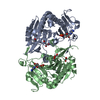
| ||||||||
|---|---|---|---|---|---|---|---|---|---|
| 1 |
| ||||||||
| Unit cell |
|
-Components
| #1: Protein | Mass: 39053.156 Da / Num. of mol.: 2 Source method: isolated from a genetically manipulated source Source: (gene. exp.)  #2: Chemical | ChemComp-ZN /   Mass: 65.409 Da / Num. of mol.: 4 / Source method: obtained synthetically / Formula: Zn Mass: 65.409 Da / Num. of mol.: 4 / Source method: obtained synthetically / Formula: Zn#3: Chemical |   Mass: 347.221 Da / Num. of mol.: 2 / Source method: obtained synthetically / Formula: C10H14N5O7P / Comment: AMP*YM Mass: 347.221 Da / Num. of mol.: 2 / Source method: obtained synthetically / Formula: C10H14N5O7P / Comment: AMP*YM#4: Chemical | ChemComp-EDO /   Mass: 62.068 Da / Num. of mol.: 6 / Source method: obtained synthetically / Formula: C2H6O2 Mass: 62.068 Da / Num. of mol.: 6 / Source method: obtained synthetically / Formula: C2H6O2#5: Water | ChemComp-HOH / |  Mass: 18.015 Da / Num. of mol.: 500 / Source method: isolated from a natural source / Formula: H2O Mass: 18.015 Da / Num. of mol.: 500 / Source method: isolated from a natural source / Formula: H2OHas protein modification | Y | |
|---|
-Experimental details
-Experiment
| Experiment | Method: X-RAY DIFFRACTION / Number of used crystals: 1 |
|---|
- Sample preparation
| Crystal | Density Matthews: 2.4 Å3/Da / Density % sol: 47.4 % |
|---|---|
| Crystal grow | Temperature: 293 K / pH: 5.5 Details: 10 MG/ML PROTEIN, 20% PEG 2000, 0.2M Sodium Chloride, 0.10M MES-Acetate, vapor diffusion, hanging drop, temperature 293K, pH 5.50 |
-Data collection
| Diffraction | Mean temperature: 100 K | |||||||||||||||||||||||||||||||||||||||||||||||||||||||||||||||||||||||||||||
|---|---|---|---|---|---|---|---|---|---|---|---|---|---|---|---|---|---|---|---|---|---|---|---|---|---|---|---|---|---|---|---|---|---|---|---|---|---|---|---|---|---|---|---|---|---|---|---|---|---|---|---|---|---|---|---|---|---|---|---|---|---|---|---|---|---|---|---|---|---|---|---|---|---|---|---|---|---|---|
| Diffraction source | Source: SYNCHROTRON / Site: APS  / Beamline: 19-BM / Wavelength: 0.97948 / Beamline: 19-BM / Wavelength: 0.97948 | |||||||||||||||||||||||||||||||||||||||||||||||||||||||||||||||||||||||||||||
| Detector | Type: APS-1 / Detector: CCD / Date: Dec 15, 2004 Details: SAGITALLY FOCUSING 2ND CRYSTAL, ROSENBAUM-ROCK VERTICAL FOCUSING MIRROR | |||||||||||||||||||||||||||||||||||||||||||||||||||||||||||||||||||||||||||||
| Radiation | Monochromator: ROSENBAUM-ROCK DOUBLE-CRYSTAL MONOCHROMATOR: WATER COOLED Protocol: SINGLE WAVELENGTH / Monochromatic (M) / Laue (L): M / Scattering type: x-ray | |||||||||||||||||||||||||||||||||||||||||||||||||||||||||||||||||||||||||||||
| Radiation wavelength | Wavelength: 0.97948 Å / Relative weight: 1 | |||||||||||||||||||||||||||||||||||||||||||||||||||||||||||||||||||||||||||||
| Reflection | Redundancy: 6.9 % / Number: 57501 / Rmerge(I) obs: 0.083 / Χ2: 1.013 / D res high: 1.83 Å / D res low: 50 Å / % possible obs: 96.5 | |||||||||||||||||||||||||||||||||||||||||||||||||||||||||||||||||||||||||||||
| Diffraction reflection shell |
| |||||||||||||||||||||||||||||||||||||||||||||||||||||||||||||||||||||||||||||
| Reflection | Resolution: 1.834→34.486 Å / Num. obs: 57501 / % possible obs: 96.5 % / Redundancy: 6.9 % / Rmerge(I) obs: 0.083 / Net I/σ(I): 15.249 | |||||||||||||||||||||||||||||||||||||||||||||||||||||||||||||||||||||||||||||
| Reflection shell | Resolution: 1.83→1.9 Å / Redundancy: 5.5 % / Rmerge(I) obs: 0.386 / Mean I/σ(I) obs: 4.543 / % possible all: 71.4 |
-Phasing
| Phasing | Method: molecular replacement | |||||||||
|---|---|---|---|---|---|---|---|---|---|---|
| Phasing MR | Rfactor: 0.394 / Cor.coef. Fo:Fc: 0.623
|
- Processing
| Software |
| ||||||||||||||||||||||||||||||||||||||||||||||||||||||||||||||||||||||||||||||||||||||||||||||||||||||||||||||||||||||||||||||||||||||||||||||||||||||||||||||||||||||||||
|---|---|---|---|---|---|---|---|---|---|---|---|---|---|---|---|---|---|---|---|---|---|---|---|---|---|---|---|---|---|---|---|---|---|---|---|---|---|---|---|---|---|---|---|---|---|---|---|---|---|---|---|---|---|---|---|---|---|---|---|---|---|---|---|---|---|---|---|---|---|---|---|---|---|---|---|---|---|---|---|---|---|---|---|---|---|---|---|---|---|---|---|---|---|---|---|---|---|---|---|---|---|---|---|---|---|---|---|---|---|---|---|---|---|---|---|---|---|---|---|---|---|---|---|---|---|---|---|---|---|---|---|---|---|---|---|---|---|---|---|---|---|---|---|---|---|---|---|---|---|---|---|---|---|---|---|---|---|---|---|---|---|---|---|---|---|---|---|---|---|---|---|
| Refinement | Method to determine structure: MOLECULAR REPLACEMENT Starting model: PDB ENTRY 1VKV 1vkv Resolution: 1.83→31.28 Å / Cor.coef. Fo:Fc: 0.956 / Cor.coef. Fo:Fc free: 0.929 / SU B: 3.115 / SU ML: 0.096 / Cross valid method: THROUGHOUT / ESU R: 0.143 / ESU R Free: 0.137 / Stereochemistry target values: MAXIMUM LIKELIHOOD
| ||||||||||||||||||||||||||||||||||||||||||||||||||||||||||||||||||||||||||||||||||||||||||||||||||||||||||||||||||||||||||||||||||||||||||||||||||||||||||||||||||||||||||
| Solvent computation | Ion probe radii: 0.8 Å / Shrinkage radii: 0.8 Å / VDW probe radii: 1.2 Å / Solvent model: MASK | ||||||||||||||||||||||||||||||||||||||||||||||||||||||||||||||||||||||||||||||||||||||||||||||||||||||||||||||||||||||||||||||||||||||||||||||||||||||||||||||||||||||||||
| Displacement parameters | Biso mean: 24.85 Å2
| ||||||||||||||||||||||||||||||||||||||||||||||||||||||||||||||||||||||||||||||||||||||||||||||||||||||||||||||||||||||||||||||||||||||||||||||||||||||||||||||||||||||||||
| Refinement step | Cycle: LAST / Resolution: 1.83→31.28 Å
| ||||||||||||||||||||||||||||||||||||||||||||||||||||||||||||||||||||||||||||||||||||||||||||||||||||||||||||||||||||||||||||||||||||||||||||||||||||||||||||||||||||||||||
| Refine LS restraints |
| ||||||||||||||||||||||||||||||||||||||||||||||||||||||||||||||||||||||||||||||||||||||||||||||||||||||||||||||||||||||||||||||||||||||||||||||||||||||||||||||||||||||||||
| LS refinement shell | Resolution: 1.83→1.88 Å / Total num. of bins used: 20
|
