 Movie
Movie Controller

[English] 日本語
 Yorodumi
- PDB-1jbq: STRUCTURE OF HUMAN CYSTATHIONINE BETA-SYNTHASE: A UNIQUE PYRIDOXA... -
+ Open data

- Basic information
| Entry | Database: PDB / ID: 1jbq | ||||||
|---|---|---|---|---|---|---|---|
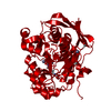
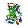
| Title | STRUCTURE OF HUMAN CYSTATHIONINE BETA-SYNTHASE: A UNIQUE PYRIDOXAL 5'-PHOSPHATE DEPENDENT HEMEPROTEIN | ||||||
 Components Components | CYSTATHIONINE BETA-SYNTHASE | ||||||
 Keywords Keywords | LYASE / Fold type II of PLP enzymes | ||||||
| Function / homology |  Function and homology information Function and homology informationCysteine formation from homocysteine / L-homocysteine catabolic process / modified amino acid binding / cystathionine beta-synthase / cystathionine beta-synthase activity / L-serine catabolic process / : / Metabolism of ingested SeMet, Sec, MeSec into H2Se / : / carbon monoxide binding ...Cysteine formation from homocysteine / L-homocysteine catabolic process / modified amino acid binding / cystathionine beta-synthase / cystathionine beta-synthase activity / L-serine catabolic process / : / Metabolism of ingested SeMet, Sec, MeSec into H2Se / : / carbon monoxide binding / cartilage development involved in endochondral bone morphogenesis / hydrogen sulfide biosynthetic process / homocysteine metabolic process / cerebellum morphogenesis / L-cysteine catabolic process / L-serine metabolic process / L-cysteine biosynthetic process / endochondral ossification / response to folic acid / transsulfuration / nitric oxide binding / : / DNA protection / S-adenosyl-L-methionine binding / nitrite reductase (NO-forming) activity / regulation of JNK cascade / superoxide metabolic process / maternal process involved in female pregnancy / blood vessel remodeling / blood vessel diameter maintenance / oxygen binding / pyridoxal phosphate binding / cellular response to hypoxia / heme binding / ubiquitin protein ligase binding / negative regulation of apoptotic process / enzyme binding / protein homodimerization activity / metal ion binding / identical protein binding / nucleus / cytoplasm / cytosol Similarity search - Function | ||||||
| Biological species |  Homo sapiens (human) Homo sapiens (human) | ||||||
| Method |  X-RAY DIFFRACTION / SYNCHROTRON / MOLECULAR REPLACEMENT / Resolution: 2.6 Å X-RAY DIFFRACTION / SYNCHROTRON / MOLECULAR REPLACEMENT / Resolution: 2.6 Å | ||||||
 Authors Authors | Meier, M. / Janosik, M. / Kery, V. / Kraus, J.P. / Burkhard, P. | ||||||
 Citation Citation | Journal: EMBO J. / Year: 2001 Title: Structure of human cystathionine beta-synthase: a unique pyridoxal 5'-phosphate-dependent heme protein. Authors: Meier, M. / Janosik, M. / Kery, V. / Kraus, J.P. / Burkhard, P. #1: Journal: Acta Crystallogr.,Sect.D / Year: 2001Title: Crystallization and preliminary X-ray diffraction analysis of the active core of human recombinant cystathionine beta-synthase: an enzyme involved in vascular disease. Authors: Janosik, M. / Meier, M. / Kery, V. / Oliveriusova, J. / Burkhard, P. / Kraus, J.P. | ||||||
| History |
|
- Structure visualization
| Structure viewer | Molecule: MolmilJmol/JSmol |
|---|
- Downloads & links
-Download
| PDBx/mmCIF format | 1jbq.cif.gz | 419 KB | Display | PDBx/mmCIF format |
|---|---|---|---|---|
| PDB format | pdb1jbq.ent.gz | 341.9 KB | Display | PDB format |
| PDBx/mmJSON format | 1jbq.json.gz | Tree view | PDBx/mmJSON format | |
| Others |  Other downloads Other downloads |
-Validation report
| Arichive directory | https://data.pdbj.org/pub/pdb/validation_reports/jb/1jbqftp://data.pdbj.org/pub/pdb/validation_reports/jb/1jbq | HTTPS FTP |
|---|
-Related structure data
| Related structure data | 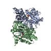 1d6sS S: Starting model for refinement |
|---|---|
| Similar structure data |


-Links
 PDBj
PDBj

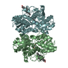
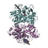
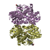
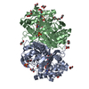
- Assembly
| Deposited unit | 
| ||||||||||
|---|---|---|---|---|---|---|---|---|---|---|---|
| 1 | 
| ||||||||||
| 2 | 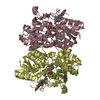
| ||||||||||
| 3 | 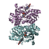
| ||||||||||
| Unit cell |
|
-Components
| #1: Protein | Mass: 47520.074 Da / Num. of mol.: 6 / Fragment: THE ACTIVE CORE / Mutation: DELETION 414-551, EXTRA 23 RESIDUES AT N-TERMINUS Source method: isolated from a genetically manipulated source Details: MINOR ISOFORM / Source: (gene. exp.) Homo sapiens (human) / Plasmid: PGEX-5X-1HCBSD414-551 / Production host:  #2: Chemical | ChemComp-HEM /   Mass: 616.487 Da / Num. of mol.: 6 / Source method: obtained synthetically / Formula: C34H32FeN4O4 Mass: 616.487 Da / Num. of mol.: 6 / Source method: obtained synthetically / Formula: C34H32FeN4O4#3: Chemical | ChemComp-PLP /   Mass: 247.142 Da / Num. of mol.: 6 / Source method: obtained synthetically / Formula: C8H10NO6P Mass: 247.142 Da / Num. of mol.: 6 / Source method: obtained synthetically / Formula: C8H10NO6P#4: Water | ChemComp-HOH / |  Mass: 18.015 Da / Num. of mol.: 154 / Source method: isolated from a natural source / Formula: H2O Mass: 18.015 Da / Num. of mol.: 154 / Source method: isolated from a natural source / Formula: H2O |
|---|
-Experimental details
-Experiment
| Experiment | Method: X-RAY DIFFRACTION / Number of used crystals: 2 |
|---|
- Sample preparation
| Crystal | Density Matthews: 2.29 Å3/Da / Density % sol: 46.19 % | ||||||||||||||||||||||||||||||||||||
|---|---|---|---|---|---|---|---|---|---|---|---|---|---|---|---|---|---|---|---|---|---|---|---|---|---|---|---|---|---|---|---|---|---|---|---|---|---|
| Crystal grow | Temperature: 293 K / Method: vapor diffusion, hanging drop / pH: 7.5 Details: PEG 1000, HEPES, pH 7.5 , VAPOR DIFFUSION, HANGING DROP at 293K | ||||||||||||||||||||||||||||||||||||
| Crystal grow | *PLUS pH: 7.4 Details: Janosik, M., (2001) Acta Crystallogr., Sect.D, 57, 289. | ||||||||||||||||||||||||||||||||||||
| Components of the solutions | *PLUS
|
-Data collection
| Diffraction |
| ||||||||||||||||||
|---|---|---|---|---|---|---|---|---|---|---|---|---|---|---|---|---|---|---|---|
| Diffraction source |
| ||||||||||||||||||
| Detector |
| ||||||||||||||||||
| Radiation |
| ||||||||||||||||||
| Radiation wavelength |
| ||||||||||||||||||
| Reflection | Resolution: 2.6→50 Å / Num. all: 87368 / Num. obs: 63971 / % possible obs: 82.4 % / Observed criterion σ(I): 2 / Redundancy: 3.3 % / Biso Wilson estimate: 29.2 Å2 / Rsym value: 0.076 / Net I/σ(I): 10 | ||||||||||||||||||
| Reflection shell | Resolution: 2.6→2.69 Å / Redundancy: 2.3 % / Mean I/σ(I) obs: 4 / Rsym value: 0.31 / % possible all: 65.1 | ||||||||||||||||||
| Reflection | *PLUS Lowest resolution: 50 Å / Rmerge(I) obs: 0.076 | ||||||||||||||||||
| Reflection shell | *PLUS % possible obs: 63.9 % / Rmerge(I) obs: 0.309 |


- Processing
| Software |
| ||||||||||||||||||||||||||||||||||||||||
|---|---|---|---|---|---|---|---|---|---|---|---|---|---|---|---|---|---|---|---|---|---|---|---|---|---|---|---|---|---|---|---|---|---|---|---|---|---|---|---|---|---|
| Refinement | Method to determine structure: MOLECULAR REPLACEMENT Starting model: PDB ENTRY 1D6S Resolution: 2.6→43.34 Å / Rfactor Rfree error: 0.005 / Data cutoff high absF: 3036819.24 / Data cutoff low absF: 0 / Isotropic thermal model: RESTRAINED / Cross valid method: THROUGHOUT / σ(F): 0 / σ(I): 0 / Stereochemistry target values: Engh & Huber Details: Residues THR 193 and GLU 201 are disordered in all chains.
| ||||||||||||||||||||||||||||||||||||||||
| Solvent computation | Solvent model: FLAT MODEL / Bsol: 29.57 Å2 / ksol: 0.341 e/Å3 | ||||||||||||||||||||||||||||||||||||||||
| Displacement parameters | Biso mean: 37.4 Å2
| ||||||||||||||||||||||||||||||||||||||||
| Refine analyze |
| ||||||||||||||||||||||||||||||||||||||||
| Refinement step | Cycle: LAST / Resolution: 2.6→43.34 Å
| ||||||||||||||||||||||||||||||||||||||||
| Refine LS restraints |
| ||||||||||||||||||||||||||||||||||||||||
| LS refinement shell | Resolution: 2.5→2.66 Å / Rfactor Rfree error: 0.033 / Total num. of bins used: 6
| ||||||||||||||||||||||||||||||||||||||||
| Xplor file |
| ||||||||||||||||||||||||||||||||||||||||
| Software | *PLUS Name: CNS / Version: 1 / Classification: refinement | ||||||||||||||||||||||||||||||||||||||||
| Refinement | *PLUS σ(F): 0 / % reflection Rfree: 5 % | ||||||||||||||||||||||||||||||||||||||||
| Solvent computation | *PLUS | ||||||||||||||||||||||||||||||||||||||||
| Displacement parameters | *PLUS Biso mean: 37.4 Å2 | ||||||||||||||||||||||||||||||||||||||||
| Refine LS restraints | *PLUS
| ||||||||||||||||||||||||||||||||||||||||
| LS refinement shell | *PLUS Rfactor Rfree: 0.358 / % reflection Rfree: 3.9 % / Rfactor Rwork: 0.3 |
