 Movie
Movie Controller

[English] 日本語
 Yorodumi
- PDB-1m0v: NMR STRUCTURE OF THE TYPE III SECRETORY DOMAIN OF YERSINIA YOPH C... -
+ Open data

- Basic information
| Entry | Database: PDB / ID: 1m0v | ||||||
|---|---|---|---|---|---|---|---|
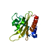
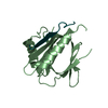
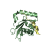
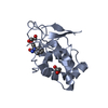
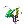
| Title | NMR STRUCTURE OF THE TYPE III SECRETORY DOMAIN OF YERSINIA YOPH COMPLEXED WITH THE SKAP-HOM PHOSPHO-PEPTIDE N-acetyl-DEpYDDPF-NH2 | ||||||
 Components Components |
| ||||||
 Keywords Keywords | HYDROLASE / high resolution structure | ||||||
| Function / homology |  Function and homology information Function and homology informationSignal regulatory protein family interactions / B cell activation / protein-tyrosine-phosphatase / protein tyrosine phosphatase activity / negative regulation of cell population proliferation / extracellular region / nucleoplasm / plasma membrane / cytoplasm / cytosol Similarity search - Function | ||||||
| Biological species |  Yersinia pseudotuberculosis (bacteria) Yersinia pseudotuberculosis (bacteria) | ||||||
| Method | SOLUTION NMR / torsion angle dynamics | ||||||
 Authors Authors | Khandelwal, P. / Keliikuli, K. / Smith, C.L. / Saper, M.A. / Zuiderweg, E.R.P. | ||||||
 Citation Citation | Journal: Biochemistry / Year: 2002 Title: Solution structure and phosphopeptide binding to the N-terminal domain of Yersinia YopH: comparison with a crystal structure Authors: Khandelwal, P. / Keliikuli, K. / Smith, C.L. / Saper, M.A. / Zuiderweg, E.R.P. #1: Journal: MOL.MICROBIOL. / Year: 2001Title: STRUCTURE OF THE TYPE III SECRETION AND SUBSTRATE-BINDING DOMAIN OF YERSINIA YOPH PHOSPHATASE Authors: SMITH, C.L. / KHANDELWAL, P. / KELIIKULI, K. / ZUIDERWEG, E.R.P. / SAPER, M.A. #2: Journal: J.BIOMOL.NMR / Year: 2001Title: 1H, 15N AND 13C ASSIGNMENTS OF THE N-TERMINAL DOMAIN OF YERSINIA OUTER PROTEIN H IN ITS APO FORM AND IN COMPLEX WITH A PHOSPHOTYROSINE PEPTIDE Authors: KHANDELWAL, P. / KELIIKULI, K. / SMITH, C.L. / SAPER, M.A. / ZUIDERWEG, E.R.P. | ||||||
| History |
|
- Structure visualization
| Structure viewer | Molecule: MolmilJmol/JSmol |
|---|
- Downloads & links
-Download
| PDBx/mmCIF format | 1m0v.cif.gz | 817.9 KB | Display | PDBx/mmCIF format |
|---|---|---|---|---|
| PDB format | pdb1m0v.ent.gz | 678.4 KB | Display | PDB format |
| PDBx/mmJSON format | 1m0v.json.gz | Tree view | PDBx/mmJSON format | |
| Others |  Other downloads Other downloads |
-Validation report
| Arichive directory | https://data.pdbj.org/pub/pdb/validation_reports/m0/1m0vftp://data.pdbj.org/pub/pdb/validation_reports/m0/1m0v | HTTPS FTP |
|---|
-Related structure data
| Similar structure data |
|---|


-Links
 PDBj
PDBj


- Assembly
| Deposited unit | 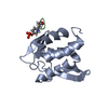
| |||||||||
|---|---|---|---|---|---|---|---|---|---|---|
| 1 |
| |||||||||
| NMR ensembles |
|
-Components
| #1: Protein | Mass: 14882.599 Da / Num. of mol.: 1 / Fragment: AMINO-TERMINAL DOMAIN (RESIDUES 1-129) Source method: isolated from a genetically manipulated source Source: (gene. exp.) Yersinia pseudotuberculosis (bacteria) / Production host: References: UniProt: p08538, UniProt: P08538*PLUS, protein-tyrosine-phosphatase |
|---|---|
| #2: Protein/peptide | Mass: 1003.879 Da / Num. of mol.: 1 / Source method: obtained synthetically Details: THIS peptide WAS CHEMICALLY synthesiZED. It is naturally found in Mus musculus (mouse). References: GenBank: 13277602, UniProt: Q3UND0*PLUS |
| Has protein modification | Y |
-Experimental details
-Experiment
| Experiment | Method: SOLUTION NMR | ||||||||||||||||||||
|---|---|---|---|---|---|---|---|---|---|---|---|---|---|---|---|---|---|---|---|---|---|
| NMR experiment |
| ||||||||||||||||||||
| NMR details | Text: 15N HSQC titrations |
- Sample preparation
| Details | Contents: 0.6 mM YopHNT U-15N,13C complexed with 0.72 mM unlabeled peptide; 50mM phosphate buffer NA Solvent system: 90% H2O/10% D2O |
|---|---|
| Sample conditions | Ionic strength: 50 mm phosphate / pH: 6.5 / Pressure: ambient / Temperature: 298 K |
| Crystal grow | *PLUS Method: other / Details: NMR |
-NMR measurement
| Radiation | Protocol: SINGLE WAVELENGTH / Monochromatic (M) / Laue (L): M | |||||||||||||||
|---|---|---|---|---|---|---|---|---|---|---|---|---|---|---|---|---|
| Radiation wavelength | Relative weight: 1 | |||||||||||||||
| NMR spectrometer |
|
- Processing
| NMR software |
| ||||||||||||||||||||
|---|---|---|---|---|---|---|---|---|---|---|---|---|---|---|---|---|---|---|---|---|---|
| Refinement | Method: torsion angle dynamics / Software ordinal: 1 Details: the structures are based on a total of 3472 restraints, 3222 are NOE-derived distance constraints, 152 dihedral angle restraints, 98 distance restraints from hydrogen bonds. | ||||||||||||||||||||
| NMR representative | Selection criteria: best ramachandran plot | ||||||||||||||||||||
| NMR ensemble | Conformer selection criteria: structures with the least restraint violations Conformers calculated total number: 360 / Conformers submitted total number: 20 |
 NMRPipe