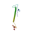
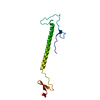
[English] 日本語
Yorodumi
- PDB-1lsl: Crystal Structure of the Thrombospondin-1 Type 1 Repeats -

+
Open data


ID or keywords:

Loading...

-
Basic information

Entry
Database: PDB / ID: 1lsl
TitleCrystal Structure of the Thrombospondin-1 Type 1 Repeats
ComponentsThrombospondin 1
KeywordsCELL ADHESION / TSP-1 / TSR
Function / homology
Function and homology information


negative regulation of antigen processing and presentation of peptide or polysaccharide antigen via MHC class II / negative regulation of nitric oxide-cGMP mediated signal transduction / negative regulation of dendritic cell antigen processing and presentation / : / collagen V binding / negative regulation of sprouting angiogenesis / chronic inflammatory response / negative regulation of endothelial cell chemotaxis / Defective B3GALTL causes PpS / O-glycosylation of TSR domain-containing proteins ...negative regulation of antigen processing and presentation of peptide or polysaccharide antigen via MHC class II / negative regulation of nitric oxide-cGMP mediated signal transduction / negative regulation of dendritic cell antigen processing and presentation / : / collagen V binding / negative regulation of sprouting angiogenesis / chronic inflammatory response / negative regulation of endothelial cell chemotaxis / Defective B3GALTL causes PpS / O-glycosylation of TSR domain-containing proteins / positive regulation of extrinsic apoptotic signaling pathway via death domain receptors / negative regulation of fibroblast growth factor receptor signaling pathway / negative regulation of long-chain fatty acid import across plasma membrane / negative regulation of blood vessel endothelial cell proliferation involved in sprouting angiogenesis / positive regulation of transforming growth factor beta1 production / engulfment of apoptotic cell / fibrinogen complex / low-density lipoprotein particle binding / negative regulation of interleukin-12 production / negative regulation of focal adhesion assembly / Signaling by PDGF / platelet alpha granule / negative regulation of plasminogen activation / fibrinogen binding / negative regulation of cell migration involved in sprouting angiogenesis / positive regulation of chemotaxis / positive regulation of macrophage activation / transforming growth factor beta binding / sprouting angiogenesis / proteoglycan binding / negative regulation of endothelial cell migration / positive regulation of fibroblast migration / negative regulation of receptor guanylyl cyclase signaling pathway / Syndecan interactions / extracellular matrix structural constituent / negative regulation of interleukin-10 production / phosphatidylserine binding / endopeptidase inhibitor activity / response to testosterone / negative regulation of endothelial cell proliferation / positive regulation of transforming growth factor beta receptor signaling pathway / positive regulation of macrophage chemotaxis / fibronectin binding / fibroblast growth factor binding / negative regulation of blood vessel endothelial cell migration / behavioral response to pain / negative regulation of fibrinolysis / negative regulation of cell-matrix adhesion / blood coagulation, fibrin clot formation / negative regulation of tumor necrosis factor production / positive regulation of phosphorylation / positive regulation of endothelial cell apoptotic process / response to magnesium ion / positive regulation of blood vessel endothelial cell migration / response to unfolded protein / Integrin cell surface interactions / response to mechanical stimulus / response to glucose / nitric oxide-cGMP-mediated signaling / response to progesterone / laminin binding / positive regulation of endothelial cell migration / positive regulation of smooth muscle cell proliferation / secretory granule / platelet alpha granule lumen / response to endoplasmic reticulum stress / negative regulation of angiogenesis / negative regulation of extrinsic apoptotic signaling pathway / sarcoplasmic reticulum / positive regulation of translation / RUNX1 regulates genes involved in megakaryocyte differentiation and platelet function / response to calcium ion / integrin binding / cellular response to growth factor stimulus / cellular response to tumor necrosis factor / positive regulation of reactive oxygen species metabolic process / positive regulation of angiogenesis / positive regulation of tumor necrosis factor production / Platelet degranulation / cell migration / heparin binding / cellular response to heat / extracellular matrix / protease binding / response to hypoxia / positive regulation of MAPK cascade / positive regulation of phosphatidylinositol 3-kinase/protein kinase B signal transduction / cell adhesion / immune response / positive regulation of cell migration / response to xenobiotic stimulus / endoplasmic reticulum lumen / inflammatory response / external side of plasma membrane / negative regulation of cell population proliferation / apoptotic process / calcium ion binding / positive regulation of cell population proliferation / negative regulation of apoptotic process / cell surface
Similarity search - Function
TSP-1 type 1 repeat / Thrombospondin type-1 (TSP1) repeat / Thrombospondin, C-terminal / Thrombospondin, type 3 repeat / Thrombospondin C-terminal region / Thrombospondin type-3 (TSP3) repeat profile. / Thrombospondin C-terminal domain profile. / Thrombospondin, type 3-like repeat / Thrombospondin type 3 repeat / TSP type-3 repeat ...TSP-1 type 1 repeat / Thrombospondin type-1 (TSP1) repeat / Thrombospondin, C-terminal / Thrombospondin, type 3 repeat / Thrombospondin C-terminal region / Thrombospondin type-3 (TSP3) repeat profile. / Thrombospondin C-terminal domain profile. / Thrombospondin, type 3-like repeat / Thrombospondin type 3 repeat / TSP type-3 repeat / : / Thrombospondin N-terminal -like domains. / von Willebrand factor type C domain / VWFC domain signature. / VWFC domain profile. / von Willebrand factor (vWF) type C domain / VWFC domain / Thrombospondin type 1 domain / Thrombospondin type-1 (TSP1) repeat superfamily / Thrombospondin type-1 (TSP1) repeat profile. / Thrombospondin type 1 repeats / Thrombospondin type-1 (TSP1) repeat / EGF-like calcium-binding domain / Calcium-binding EGF-like domain / Single Sheet / Epidermal growth factor-like domain. / EGF-like domain profile. / EGF-like domain signature 2. / EGF-like domain / Concanavalin A-like lectin/glucanase domain superfamily / Mainly Beta
Similarity search - Domain/homology
alpha-L-fucopyranose / beta-L-fucopyranose / Thrombospondin-1
Similarity search - Component
Biological speciesHomo sapiens (human)
MethodX-RAY DIFFRACTION / SYNCHROTRON / MAD / Resolution: 1.9 Å
AuthorsTan, K. / Duquette, M. / Liu, J. / Dong, Y. / Zhang, R. / Joachimiak, A. / Lawler, J. / Wang, J.-H.
CitationJournal: J.Cell Biol. / Year: 2002
Title: Crystal structure of the TSP-1 type 1 repeats: a novel layered fold and its biological implication.
Authors: Tan, K. / Duquette, M. / Liu, J.H. / Dong, Y. / Zhang, R. / Joachimiak, A. / Lawler, J. / Wang, J.H.
History
DepositionMay 17, 2002Deposition site: RCSB / Processing site: RCSB
Revision 1.0Dec 18, 2002Provider: repository / Type: Initial release
Revision 1.1Apr 28, 2008Group: Version format compliance
Revision 1.2Jul 13, 2011Group: Version format compliance
Revision 1.3Jul 29, 2020Group: Advisory / Data collection ...Advisory / Data collection / Derived calculations / Structure summary
Category: chem_comp / database_PDB_caveat ...chem_comp / database_PDB_caveat / entity / pdbx_chem_comp_identifier / pdbx_entity_nonpoly / struct_conn / struct_site / struct_site_gen
Item: _chem_comp.name / _chem_comp.type ..._chem_comp.name / _chem_comp.type / _entity.pdbx_description / _pdbx_entity_nonpoly.name / _struct_conn.pdbx_leaving_atom_flag / _struct_conn.pdbx_role
Description: Carbohydrate remediation / Provider: repository / Type: Remediation
Revision 1.4Oct 30, 2024Group: Data collection / Database references / Structure summary
Category: chem_comp / chem_comp_atom ...chem_comp / chem_comp_atom / chem_comp_bond / database_2 / pdbx_entry_details / pdbx_modification_feature
Item: _chem_comp.pdbx_synonyms / _database_2.pdbx_DOI / _database_2.pdbx_database_accession

-
Structure visualization

Structure viewerMolecule:
MolmilJmol/JSmol

Downloads & links

-
Assembly

Deposited unit
A: Thrombospondin 1
hetero molecules


Theoretical massNumber of molelcules
Total (without water)12,5053
Polymers12,1771
Non-polymers3282
Water2,774154
1


  • Idetical with deposited unit
  • defined by author
TypeNameSymmetry operationNumber
identity operation1_555x,y,z1
Unit cell
Length a, b, c (Å)67.63, 84.41, 37.17
Angle α, β, γ (deg.)90, 108.67, 90
Int Tables number5
Space group name H-MC121

-
Components

#1: Protein Thrombospondin 1


Mass: 12176.628 Da / Num. of mol.: 1 / Fragment: TSP Type 1 Repeats 2 and 3 (residues 434-546)
Source method: isolated from a genetically manipulated source
Source: (gene. exp.) Homo sapiens (human) / Gene: THBS1 / Plasmid: PMT-BIP-V5-HIS A / Genus (production host): Drosophila / Cell (production host): S2 cells / Production host: Drosophila (fruit flies) / References: UniProt: P07996
#2: Sugar ChemComp-FUL / beta-L-fucopyranose / beta-L-fucose / 6-deoxy-beta-L-galactopyranose / L-fucose / fucose / 6-DEOXY-BETA-L-GALACTOSE


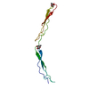
Type: L-saccharide, beta linking / Mass: 164.156 Da / Num. of mol.: 1
Source method: isolated from a genetically manipulated source
Formula: C6H12O5
IdentifierTypeProgram
LFucpbCONDENSED IUPAC CARBOHYDRATE SYMBOLGMML 1.0
b-L-fucopyranoseCOMMON NAMEGMML 1.0
b-L-FucpIUPAC CARBOHYDRATE SYMBOLPDB-CARE 1.0
FucSNFG CARBOHYDRATE SYMBOLGMML 1.0
#3: Sugar ChemComp-FUC / alpha-L-fucopyranose / alpha-L-fucose / 6-deoxy-alpha-L-galactopyranose / L-fucose / fucose


Type: L-saccharide, alpha linking / Mass: 164.156 Da / Num. of mol.: 1
Source method: isolated from a genetically manipulated source
Formula: C6H12O5
IdentifierTypeProgram
LFucpaCONDENSED IUPAC CARBOHYDRATE SYMBOLGMML 1.0
a-L-fucopyranoseCOMMON NAMEGMML 1.0
a-L-FucpIUPAC CARBOHYDRATE SYMBOLPDB-CARE 1.0
FucSNFG CARBOHYDRATE SYMBOLGMML 1.0
#4: Water ChemComp-HOH / water


Mass: 18.015 Da / Num. of mol.: 154 / Source method: isolated from a natural source / Formula: H2O
Has protein modificationY

-
Experimental details

-
Experiment

ExperimentMethod: X-RAY DIFFRACTION / Number of used crystals: 2

-
Sample preparation

CrystalDensity Matthews: 3.56 Å3/Da / Density % sol: 65.2 %
Crystal growTemperature: 298 K / Method: vapor diffusion, hanging drop / pH: 5.5
Details: 0.5 M sodium potassium tartrate and 0.1 M sodium acetate, pH 5.5, VAPOR DIFFUSION, HANGING DROP, temperature 298K
Crystal grow
*PLUS
Method: vapor diffusion, hanging drop
Components of the solutions
*PLUS
IDConc.Common nameCrystal-IDSol-IDDetails (eV)
10.5 Msodium potassium tartrate1reservoir
20.1 Msodium acetate1reservoirpH5.5

-
Data collection

Diffraction
IDMean temperature (K)Crystal-ID
11001
21001
Diffraction source
SourceSiteBeamlineIDWavelength (Å)
SYNCHROTRONAPS 19-ID11.00803, 1.00808, 1.0596
SYNCHROTRONAPS 19-ID21.1
Detector
TypeIDDetectorDate
ADSC QUANTUM 41CCDDec 17, 2000
ADSC QUANTUM 42CCDDec 17, 2000
Radiation
IDMonochromatorProtocolMonochromatic (M) / Laue (L)Scattering typeWavelength-ID
1GraphiteMADMx-ray1
2GraphiteSINGLE WAVELENGTHMx-ray1
Radiation wavelength
IDWavelength (Å)Relative weight
11.008031
21.008081
31.05961
41.11
ReflectionResolution: 1.9→20 Å / Num. obs: 15123 / % possible obs: 99.8 % / Observed criterion σ(I): 0 / Biso Wilson estimate: 26.4 Å2 / Rmerge(I) obs: 0.075 / Net I/σ(I): 14.6
Reflection shellResolution: 1.9→1.97 Å / Rmerge(I) obs: 0.469 / Mean I/σ(I) obs: 3.8 / % possible all: 99.7
Reflection
*PLUS
Highest resolution: 1.9 Å / Lowest resolution: 20 Å / Num. obs: 15485 / Num. measured all: 162978
Reflection shell
*PLUS
% possible obs: 99.7 %

-
Processing

Software
NameVersionClassification
HKL-2000data collection
HKL-2000data reduction
MLPHAREphasing
X-PLOR3.851refinement
HKL-2000data scaling
RefinementMethod to determine structure: MAD / Resolution: 1.9→20 Å / σ(F): 0 / Stereochemistry target values: Engh & Huber
RfactorNum. reflection% reflectionSelection details
Rfree0.282 1533 10 %Random
Rwork0.238 ---
all0.235 15123 --
obs0.235 15123 99.8 %-
Displacement parametersBiso mean: 42.28 Å2
Refine analyze
FreeObs
Luzzati coordinate error0.345 Å0.321 Å
Luzzati d res low-3 Å
Luzzati sigma a0.345 Å0.321 Å
Refinement stepCycle: LAST / Resolution: 1.9→20 Å
ProteinNucleic acidLigandSolventTotal
Num. atoms844 0 20 154 1018
Refine LS restraints
Refine-IDTypeDev ideal
X-RAY DIFFRACTIONx_bond_d0.008
X-RAY DIFFRACTIONx_angle_deg1.631
X-RAY DIFFRACTIONx_dihedral_angle_d27.854
X-RAY DIFFRACTIONx_improper_angle_d0.825
LS refinement shellResolution: 1.9→1.99 Å / Total num. of bins used: 8
RfactorNum. reflection
Rfree0.457 168
Rwork0.439 1545
obs-1545
Refinement
*PLUS
Lowest resolution: 20 Å / % reflection Rfree: 10 %
Solvent computation
*PLUS
Displacement parameters
*PLUS
Refine LS restraints
*PLUS
Refine-IDTypeDev ideal
X-RAY DIFFRACTIONx_dihedral_angle_d
X-RAY DIFFRACTIONx_dihedral_angle_deg27.854
X-RAY DIFFRACTIONx_improper_angle_d
X-RAY DIFFRACTIONx_improper_angle_deg0.825

+
About Yorodumi

-
News

-
Feb 9, 2022. New format data for meta-information of EMDB entries

New format data for meta-information of EMDB entries

  • Version 3 of the EMDB header file is now the official format.
  • The previous official version 1.9 will be removed from the archive.

Related info.:EMDB header

External links:wwPDB to switch to version 3 of the EMDB data model

-
Aug 12, 2020. Covid-19 info

Covid-19 info

URL: https://pdbj.org/emnavi/covid19.php

New page: Covid-19 featured information page in EM Navigator.

Related info.:Covid-19 info / Mar 5, 2020. Novel coronavirus structure data

+
Mar 5, 2020. Novel coronavirus structure data

Novel coronavirus structure data

Related info.:Yorodumi Speices / Aug 12, 2020. Covid-19 info

External links:COVID-19 featured content - PDBj / Molecule of the Month (242):Coronavirus Proteases

+
Jan 31, 2019. EMDB accession codes are about to change! (news from PDBe EMDB page)

EMDB accession codes are about to change! (news from PDBe EMDB page)

  • The allocation of 4 digits for EMDB accession codes will soon come to an end. Whilst these codes will remain in use, new EMDB accession codes will include an additional digit and will expand incrementally as the available range of codes is exhausted. The current 4-digit format prefixed with “EMD-” (i.e. EMD-XXXX) will advance to a 5-digit format (i.e. EMD-XXXXX), and so on. It is currently estimated that the 4-digit codes will be depleted around Spring 2019, at which point the 5-digit format will come into force.
  • The EM Navigator/Yorodumi systems omit the EMD- prefix.

Related info.:Q: What is EMD? / ID/Accession-code notation in Yorodumi/EM Navigator

External links:EMDB Accession Codes are Changing Soon! / Contact to PDBj

+
Jul 12, 2017. Major update of PDB

Major update of PDB

  • wwPDB released updated PDB data conforming to the new PDBx/mmCIF dictionary.
  • This is a major update changing the version number from 4 to 5, and with Remediation, in which all the entries are updated.
  • In this update, many items about electron microscopy experimental information are reorganized (e.g. em_software).
  • Now, EM Navigator and Yorodumi are based on the updated data.

External links:wwPDB Remediation / Enriched Model Files Conforming to OneDep Data Standards Now Available in the PDB FTP Archive

-
Yorodumi

Thousand views of thousand structures

  • Yorodumi is a browser for structure data from EMDB, PDB, SASBDB, etc.
  • This page is also the successor to EM Navigator detail page, and also detail information page/front-end page for Omokage search.
  • The word "yorodu" (or yorozu) is an old Japanese word meaning "ten thousand". "mi" (miru) is to see.

Related info.:EMDB / PDB / SASBDB / Comparison of 3 databanks / Yorodumi Search / Aug 31, 2016. New EM Navigator & Yorodumi / Yorodumi Papers / Jmol/JSmol / Function and homology information / Changes in new EM Navigator and Yorodumi

Read more