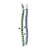
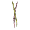
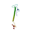
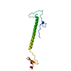
Entry Database : PDB / ID : 3r6bTitle Crystal Structure of Thrombospondin-1 TSR Domains 2 and 3 Thrombospondin-1 Keywords / / / Function / homology Function Domain/homology Component
/ / / / / / / / / / / / / / / / / / / / / / / / / / / / / / / / / / / / / / / / / / / / / / / / / / / / / / / / / / / / / / / / / / / / / / / / / / / / / / / / / / / / / / / / / / / / / / / / / / / / / / / / / / / / / / / / / / / / / / / / / / / / / / / / / / / / / / / / / / / / / / / / / / / Biological species Homo sapiens (human)Method / / Resolution : 2.4 Å Authors Page, R.C. / Klenotic, P.A. / Misra, S. / Silverstein, R.L. Journal : Protein Expr.Purif. / Year : 2011Title : Expression, purification and structural characterization of functionally replete thrombospondin-1 type 1 repeats in a bacterial expression system.Authors : Klenotic, P.A. / Page, R.C. / Misra, S. / Silverstein, R.L. History Deposition Mar 21, 2011 Deposition site / Processing site Revision 1.0 Aug 10, 2011 Provider / Type Revision 1.1 Aug 17, 2011 Group Revision 1.2 Nov 16, 2011 Group Revision 1.3 Sep 13, 2023 Group Data collection / Database references ... Data collection / Database references / Derived calculations / Refinement description Category chem_comp_atom / chem_comp_bond ... chem_comp_atom / chem_comp_bond / database_2 / pdbx_initial_refinement_model / struct_ref_seq_dif / struct_site Item _database_2.pdbx_DOI / _database_2.pdbx_database_accession ... _database_2.pdbx_DOI / _database_2.pdbx_database_accession / _struct_ref_seq_dif.details / _struct_site.pdbx_auth_asym_id / _struct_site.pdbx_auth_comp_id / _struct_site.pdbx_auth_seq_id Revision 1.4 Nov 20, 2024 Group / Category / pdbx_modification_feature
Show all Show less
 Movie
Movie Controller


 Open data
 Basic information
 Components
 Keywords
 Function and homology information
 Homo sapiens (human)
 X-RAY DIFFRACTION /
  Authors
 Citation
 Structure visualization
 Downloads & links
 Other downloads
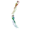


 PDBj
PDBj


 Assembly


 Mass: 62.068 Da / Num. of mol.: 8 / Source method: obtained synthetically / Formula: C2H6O2
 Mass: 18.015 Da / Num. of mol.: 94 / Source method: isolated from a natural source / Formula: H2O
 Sample preparation
 Processing