 Movie
Movie Controller


+ Open data

- Basic information
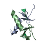
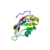
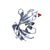
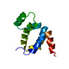
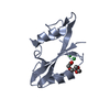
| Entry | Database: PDB / ID: 1ln4 | ||||||
|---|---|---|---|---|---|---|---|
| Title | CRYSTAL STRUCTURE OF E. COLI YHBY | ||||||
 Components Components | Hypothetical protein yhbY | ||||||
 Keywords Keywords | RNA BINDING PROTEIN / PUTATIVE RNA-BINDING PROTEIN / PUTATIVE TRANSLATION FACTOR | ||||||
| Function / homology |  Function and homology information Function and homology informationrRNA 5'-end processing / preribosome binding / ribosomal small subunit assembly / ribosomal large subunit assembly / RNA binding / cytosol Similarity search - Function | ||||||
| Biological species |  | ||||||
| Method |  X-RAY DIFFRACTION / SYNCHROTRON / MAD / Resolution: 1.5 Å X-RAY DIFFRACTION / SYNCHROTRON / MAD / Resolution: 1.5 Å | ||||||
 Authors Authors | Ostheimer, G.J. / Barkan, A. / Matthews, B.W. | ||||||
 Citation Citation | Journal: Structure / Year: 2002 Title: Crystal structure of E. coli YhbY: a representative of a novel class of RNA binding proteins Authors: Ostheimer, G.J. / Barkan, A. / Matthews, B.W. | ||||||
| History |
|
- Structure visualization
| Structure viewer | Molecule: MolmilJmol/JSmol |
|---|
- Downloads & links
-Download
| PDBx/mmCIF format | 1ln4.cif.gz | 32.5 KB | Display | PDBx/mmCIF format |
|---|---|---|---|---|
| PDB format | pdb1ln4.ent.gz | 21.9 KB | Display | PDB format |
| PDBx/mmJSON format | 1ln4.json.gz | Tree view | PDBx/mmJSON format | |
| Others |  Other downloads Other downloads |
-Validation report
| Arichive directory | https://data.pdbj.org/pub/pdb/validation_reports/ln/1ln4ftp://data.pdbj.org/pub/pdb/validation_reports/ln/1ln4 | HTTPS FTP |
|---|
-Related structure data
| Similar structure data |
|---|


-Links
 PDBj
PDBj- Assembly
| Deposited unit | 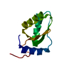
| ||||||||
|---|---|---|---|---|---|---|---|---|---|
| 1 |
| ||||||||
| Unit cell |
|
-Components
| #1: Protein | Mass: 11717.667 Da / Num. of mol.: 1 / Mutation: N2D,R97L Source method: isolated from a genetically manipulated source Source: (gene. exp.) |
|---|---|
| #2: Water | ChemComp-HOH /  Mass: 18.015 Da / Num. of mol.: 107 / Source method: isolated from a natural source / Formula: H2O Mass: 18.015 Da / Num. of mol.: 107 / Source method: isolated from a natural source / Formula: H2O |
-Experimental details
-Experiment
| Experiment | Method: X-RAY DIFFRACTION / Number of used crystals: 1 |
|---|
- Sample preparation
| Crystal | Density Matthews: 2.87 Å3/Da / Density % sol: 57.12 % | |||||||||||||||||||||||||||||||||||||||||||||||||||||||||||||||
|---|---|---|---|---|---|---|---|---|---|---|---|---|---|---|---|---|---|---|---|---|---|---|---|---|---|---|---|---|---|---|---|---|---|---|---|---|---|---|---|---|---|---|---|---|---|---|---|---|---|---|---|---|---|---|---|---|---|---|---|---|---|---|---|---|
| Crystal grow | Temperature: 298 K / Method: vapor diffusion, hanging drop / pH: 8.5 Details: PEG 200, sodium chloride, tris, pH 8.5, VAPOR DIFFUSION, HANGING DROP, temperature 298K | |||||||||||||||||||||||||||||||||||||||||||||||||||||||||||||||
| Crystal grow | *PLUS pH: 7 | |||||||||||||||||||||||||||||||||||||||||||||||||||||||||||||||
| Components of the solutions | *PLUS
|
-Data collection
| Diffraction |
| ||||||||||||||||||
|---|---|---|---|---|---|---|---|---|---|---|---|---|---|---|---|---|---|---|---|
| Diffraction source |
| ||||||||||||||||||
| Detector |
| ||||||||||||||||||
| Radiation |
| ||||||||||||||||||
| Radiation wavelength |
| ||||||||||||||||||
| Reflection | Resolution: 1.5→20 Å / Num. all: 22101 / Num. obs: 22089 / % possible obs: 99.9 % / Observed criterion σ(F): 0 / Observed criterion σ(I): 0 / Redundancy: 10.6 % / Biso Wilson estimate: 14 Å2 / Rmerge(I) obs: 0.074 / Rsym value: 0.074 / Net I/σ(I): 4.5 | ||||||||||||||||||
| Reflection shell | Resolution: 1.5→1.58 Å / Redundancy: 10.6 % / Rmerge(I) obs: 0.139 / Mean I/σ(I) obs: 4.5 / Num. unique all: 3170 / Rsym value: 0.139 / % possible all: 99.9 | ||||||||||||||||||
| Reflection | *PLUS Num. obs: 22061 / % possible obs: 100 % | ||||||||||||||||||
| Reflection shell | *PLUS % possible obs: 99.9 % |

- Processing
| Software |
| ||||||||||||||||||||||||||||||||||||||||||||||||||
|---|---|---|---|---|---|---|---|---|---|---|---|---|---|---|---|---|---|---|---|---|---|---|---|---|---|---|---|---|---|---|---|---|---|---|---|---|---|---|---|---|---|---|---|---|---|---|---|---|---|---|---|
| Refinement | Method to determine structure: MAD / Resolution: 1.5→20 Å / Isotropic thermal model: TNT BCORREL V1.0 / Cross valid method: THROUGHOUT / σ(F): 0 / σ(I): 0 / Stereochemistry target values: TNT GEOMETRY V1.0 Details: PHASES WERE DETERMINED USING A 3-WAVELENGTH MAD DATASET COLLECTED ON A SINGLE CRYSTAL. THE STRUCTURE WAS REFINED AGAINST AN ADDITIONAL MONOCHROMATIC DATASET THAT WAS COLLECTED USING THE SAME CRYSTAL.
| ||||||||||||||||||||||||||||||||||||||||||||||||||
| Solvent computation | Bsol: 207.501 Å2 / ksol: 0.82848 e/Å3 | ||||||||||||||||||||||||||||||||||||||||||||||||||
| Refinement step | Cycle: LAST / Resolution: 1.5→20 Å
| ||||||||||||||||||||||||||||||||||||||||||||||||||
| Refine LS restraints |
| ||||||||||||||||||||||||||||||||||||||||||||||||||
| Refinement | *PLUS Num. reflection obs: 21928 / % reflection Rfree: 5 % | ||||||||||||||||||||||||||||||||||||||||||||||||||
| Solvent computation | *PLUS | ||||||||||||||||||||||||||||||||||||||||||||||||||
| Displacement parameters | *PLUS | ||||||||||||||||||||||||||||||||||||||||||||||||||
| Refine LS restraints | *PLUS
|
