 Movie
Movie Controller


+ Open data

- Basic information
| Entry | Database: PDB / ID: 1li0 | ||||||
|---|---|---|---|---|---|---|---|
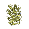
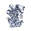
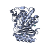
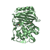
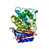
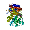
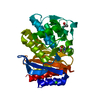
| Title | Crystal structure of TEM-32 beta-Lactamase at 1.6 Angstrom | ||||||
 Components Components | Class A beta-Lactamase- TEM-32 | ||||||
 Keywords Keywords | HYDROLASE / beta-Lactamase / antibiotic resistance / TEM-32 | ||||||
| Function / homology |  Function and homology information Function and homology informationAntimicrobial resistance / beta-lactam antibiotic catabolic process / beta-lactamase activity / beta-lactamase / response to antibiotic Similarity search - Function | ||||||
| Biological species |  | ||||||
| Method |  X-RAY DIFFRACTION / SYNCHROTRON / MOLECULAR REPLACEMENT / Resolution: 1.61 Å X-RAY DIFFRACTION / SYNCHROTRON / MOLECULAR REPLACEMENT / Resolution: 1.61 Å | ||||||
 Authors Authors | Wang, X. / Minasov, G. / Shoichet, B.K. | ||||||
 Citation Citation | Journal: J.Biol.Chem. / Year: 2002 Title: The structural bases of antibiotic resistance in the clinically derived mutant beta-lactamases TEM-30, TEM-32, and TEM-34. Authors: Wang, X. / Minasov, G. / Shoichet, B.K. | ||||||
| History |
|
- Structure visualization
| Structure viewer | Molecule: MolmilJmol/JSmol |
|---|
- Downloads & links
-Download
| PDBx/mmCIF format | 1li0.cif.gz | 74.8 KB | Display | PDBx/mmCIF format |
|---|---|---|---|---|
| PDB format | pdb1li0.ent.gz | 54.1 KB | Display | PDB format |
| PDBx/mmJSON format | 1li0.json.gz | Tree view | PDBx/mmJSON format | |
| Others |  Other downloads Other downloads |
-Validation report
| Arichive directory | https://data.pdbj.org/pub/pdb/validation_reports/li/1li0ftp://data.pdbj.org/pub/pdb/validation_reports/li/1li0 | HTTPS FTP |
|---|
-Related structure data
| Related structure data |  1lhyC  1li9C  1jwpS C: citing same article ( S: Starting model for refinement |
|---|---|
| Similar structure data |


-Links
 PDBj
PDBj


- Assembly
| Deposited unit | 
| ||||||||
|---|---|---|---|---|---|---|---|---|---|
| 1 |
| ||||||||
| Unit cell |
|
-Components
| #1: Protein | Mass: 28893.865 Da / Num. of mol.: 1 / Mutation: M69I, M182T Source method: isolated from a genetically manipulated source Source: (gene. exp.) | ||||
|---|---|---|---|---|---|
| #2: Chemical | ChemComp-BCT /   Mass: 61.017 Da / Num. of mol.: 1 / Source method: obtained synthetically / Formula: CHO3 / Comment: pH buffer*YM Mass: 61.017 Da / Num. of mol.: 1 / Source method: obtained synthetically / Formula: CHO3 / Comment: pH buffer*YM | ||||
| #3: Chemical |   Mass: 39.098 Da / Num. of mol.: 2 / Source method: obtained synthetically / Formula: K Mass: 39.098 Da / Num. of mol.: 2 / Source method: obtained synthetically / Formula: K#4: Water | ChemComp-HOH / |  Mass: 18.015 Da / Num. of mol.: 328 / Source method: isolated from a natural source / Formula: H2O Mass: 18.015 Da / Num. of mol.: 328 / Source method: isolated from a natural source / Formula: H2OHas protein modification | Y | |
-Experimental details
-Experiment
| Experiment | Method: X-RAY DIFFRACTION / Number of used crystals: 1 |
|---|
- Sample preparation
| Crystal | Density Matthews: 1.91 Å3/Da / Density % sol: 35.72 % | ||||||||||||||||||||||||
|---|---|---|---|---|---|---|---|---|---|---|---|---|---|---|---|---|---|---|---|---|---|---|---|---|---|
| Crystal grow | Temperature: 295 K / Method: vapor diffusion, hanging drop / pH: 8 Details: sodium-potassium buffer, pH 8.0, VAPOR DIFFUSION, HANGING DROP, temperature 295.0K | ||||||||||||||||||||||||
| Crystal grow | *PLUS Details: used seeding | ||||||||||||||||||||||||
| Components of the solutions | *PLUS
|
-Data collection
| Diffraction | Mean temperature: 100 K |
|---|---|
| Diffraction source | Source: SYNCHROTRON / Site: APS  / Beamline: 5ID-B / Wavelength: 1 Å / Beamline: 5ID-B / Wavelength: 1 Å |
| Detector | Type: MARRESEARCH / Detector: CCD / Date: Dec 19, 2001 / Details: Mirrors |
| Radiation | Monochromator: Graphite / Protocol: SINGLE WAVELENGTH / Monochromatic (M) / Laue (L): M / Scattering type: x-ray |
| Radiation wavelength | Wavelength: 1 Å / Relative weight: 1 |
| Reflection | Resolution: 1.61→17 Å / Num. all: 29385 / Num. obs: 29385 / % possible obs: 99.2 % / Observed criterion σ(I): -3 / Redundancy: 6.2 % / Biso Wilson estimate: 30.2 Å2 / Rmerge(I) obs: 0.061 / Net I/σ(I): 24.6 |
| Reflection shell | Resolution: 1.61→1.67 Å / Redundancy: 5.6 % / Rmerge(I) obs: 0.273 / Mean I/σ(I) obs: 6.1 / Num. unique all: 2931 / % possible all: 99.8 |
| Reflection | *PLUS Lowest resolution: 17 Å / Num. measured all: 181694 |
| Reflection shell | *PLUS % possible obs: 99.8 % |
- Processing
| Software |
| |||||||||||||||||||||||||
|---|---|---|---|---|---|---|---|---|---|---|---|---|---|---|---|---|---|---|---|---|---|---|---|---|---|---|
| Refinement | Method to determine structure: MOLECULAR REPLACEMENT Starting model: PDB ID 1JWP Resolution: 1.61→17 Å / Isotropic thermal model: Isotropic / Cross valid method: THROUGHOUT / σ(F): 0 / σ(I): 0 / Stereochemistry target values: Engh & Huber Details: Crystallographic conjugate gradient minimization refinement using maximum likelihood target for amplitudes
| |||||||||||||||||||||||||
| Displacement parameters | Biso mean: 25.4 Å2
| |||||||||||||||||||||||||
| Refine analyze |
| |||||||||||||||||||||||||
| Refinement step | Cycle: LAST / Resolution: 1.61→17 Å
| |||||||||||||||||||||||||
| Refine LS restraints |
| |||||||||||||||||||||||||
| LS refinement shell | Resolution: 1.61→1.67 Å
| |||||||||||||||||||||||||
| Refinement | *PLUS % reflection Rfree: 10 % | |||||||||||||||||||||||||
| Solvent computation | *PLUS | |||||||||||||||||||||||||
| Displacement parameters | *PLUS | |||||||||||||||||||||||||
| Refine LS restraints | *PLUS
|
