 Movie
Movie Controller

[English] 日本語
 Yorodumi
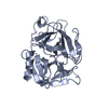
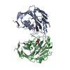
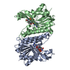
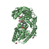
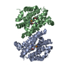
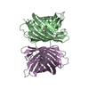
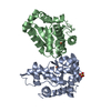
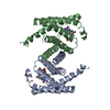
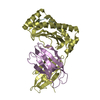
- PDB-1l4z: X-RAY CRYSTAL STRUCTURE OF THE COMPLEX OF MICROPLASMINOGEN WITH A... -
+ Open data

- Basic information
| Entry | Database: PDB / ID: 1l4z | ||||||
|---|---|---|---|---|---|---|---|
| Title | X-RAY CRYSTAL STRUCTURE OF THE COMPLEX OF MICROPLASMINOGEN WITH ALPHA DOMAIN OF STREPTOKINASE IN THE PRESENCE CADMIUM IONS | ||||||
 Components Components |
| ||||||
 Keywords Keywords | HYDROLASE/BLOOD CLOTTING / plasminogen / streptokinase / protein complex / HYDROLASE-BLOOD CLOTTING COMPLEX | ||||||
| Function / homology |  Function and homology information Function and homology informationplasmin / trans-synaptic signaling by BDNF, modulating synaptic transmission / trophoblast giant cell differentiation / tissue remodeling / tissue regeneration / Signaling by PDGF / mononuclear cell migration / positive regulation of fibrinolysis / negative regulation of cell-cell adhesion mediated by cadherin / protein antigen binding ...plasmin / trans-synaptic signaling by BDNF, modulating synaptic transmission / trophoblast giant cell differentiation / tissue remodeling / tissue regeneration / Signaling by PDGF / mononuclear cell migration / positive regulation of fibrinolysis / negative regulation of cell-cell adhesion mediated by cadherin / protein antigen binding / Dissolution of Fibrin Clot / myoblast differentiation / labyrinthine layer blood vessel development / biological process involved in interaction with symbiont / muscle cell cellular homeostasis / Activation of Matrix Metalloproteinases / apolipoprotein binding / extracellular matrix disassembly / negative regulation of fibrinolysis / negative regulation of cell-substrate adhesion / positive regulation of blood vessel endothelial cell migration / fibrinolysis / Degradation of the extracellular matrix / serine-type peptidase activity / platelet alpha granule lumen / protein processing / kinase binding / Schaffer collateral - CA1 synapse / Regulation of Insulin-like Growth Factor (IGF) transport and uptake by Insulin-like Growth Factor Binding Proteins (IGFBPs) / blood coagulation / Platelet degranulation / protein-folding chaperone binding / : / protease binding / endopeptidase activity / blood microparticle / signaling receptor binding / protein domain specific binding / negative regulation of cell population proliferation / external side of plasma membrane / serine-type endopeptidase activity / glutamatergic synapse / enzyme binding / cell surface / proteolysis / extracellular space / extracellular exosome / extracellular region / plasma membrane Similarity search - Function | ||||||
| Biological species |  Homo sapiens (human) Homo sapiens (human) Streptococcus dysgalactiae subsp. equisimilis (bacteria) Streptococcus dysgalactiae subsp. equisimilis (bacteria) | ||||||
| Method |  X-RAY DIFFRACTION / MOLECULAR REPLACEMENT / Resolution: 2.8 Å X-RAY DIFFRACTION / MOLECULAR REPLACEMENT / Resolution: 2.8 Å | ||||||
 Authors Authors | Wakeham, N. / Terzyan, S. / Zhai, P. / Loy, J.A. / Tang, J. / Zhang, X.C. | ||||||
 Citation Citation | Journal: PROTEIN ENG. / Year: 2002 Title: Effects of deletion of streptokinase residues 48-59 on plasminogen activation. Authors: Wakeham, N. / Terzyan, S. / Zhai, P. / Loy, J.A. / Tang, J. / Zhang, X.C. | ||||||
| History |
|
- Structure visualization
| Structure viewer | Molecule: MolmilJmol/JSmol |
|---|
- Downloads & links
-Download
| PDBx/mmCIF format | 1l4z.cif.gz | 88.6 KB | Display | PDBx/mmCIF format |
|---|---|---|---|---|
| PDB format | pdb1l4z.ent.gz | 66.2 KB | Display | PDB format |
| PDBx/mmJSON format | 1l4z.json.gz | Tree view | PDBx/mmJSON format | |
| Others |  Other downloads Other downloads |
-Validation report
| Summary document | 1l4z_validation.pdf.gz | 441.7 KB | Display | wwPDB validaton report |
|---|---|---|---|---|
| Full document | 1l4z_full_validation.pdf.gz | 454 KB | Display | |
| Data in XML | 1l4z_validation.xml.gz | 17.6 KB | Display | |
| Data in CIF | 1l4z_validation.cif.gz | 23.7 KB | Display | |
| Arichive directory | https://data.pdbj.org/pub/pdb/validation_reports/l4/1l4zftp://data.pdbj.org/pub/pdb/validation_reports/l4/1l4z | HTTPS FTP |
-Related structure data
| Related structure data |  1l4dC 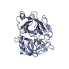 1ddjS C: citing same article ( S: Starting model for refinement |
|---|---|
| Similar structure data |


-Links
 PDBj
PDBj


- Assembly
| Deposited unit | 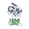
| ||||||||
|---|---|---|---|---|---|---|---|---|---|
| 1 |
| ||||||||
| 2 | 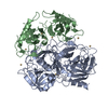
| ||||||||
| Unit cell |
| ||||||||
| Details | One molecule of microplasminogen and one molecule of streptokinase N terminal alpha domain in assymetric unit. |
-Components
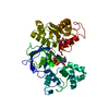
| #1: Protein | Mass: 27177.248 Da / Num. of mol.: 1 / Fragment: Catalytic domain, Residues 544-791 / Mutation: S741A Source method: isolated from a genetically manipulated source Source: (gene. exp.) Homo sapiens (human) / Production host: | ||||
|---|---|---|---|---|---|
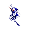
| #2: Protein | Mass: 14907.698 Da / Num. of mol.: 1 / Fragment: N terminal alpha domain, Residues 0-147 / Mutation: Q5E, W6A Source method: isolated from a genetically manipulated source Source: (gene. exp.) Streptococcus dysgalactiae subsp. equisimilis (bacteria)Species: Streptococcus dysgalactiae / Strain: subsp. equisimilis / Species (production host): Escherichia coli / Production host: | ||||
| #3: Chemical | ChemComp-CD /   Mass: 112.411 Da / Num. of mol.: 6 / Source method: obtained synthetically / Formula: Cd Mass: 112.411 Da / Num. of mol.: 6 / Source method: obtained synthetically / Formula: Cd#4: Water | ChemComp-HOH / |  Mass: 18.015 Da / Num. of mol.: 70 / Source method: isolated from a natural source / Formula: H2O Mass: 18.015 Da / Num. of mol.: 70 / Source method: isolated from a natural source / Formula: H2OHas protein modification | Y | |
-Experimental details
-Experiment
| Experiment | Method: X-RAY DIFFRACTION / Number of used crystals: 1 |
|---|
- Sample preparation
| Crystal | Density Matthews: 3.24 Å3/Da / Density % sol: 55.6 % | ||||||||||||||||||||||||||||||||||||||||||||||||||||||||||||||||||
|---|---|---|---|---|---|---|---|---|---|---|---|---|---|---|---|---|---|---|---|---|---|---|---|---|---|---|---|---|---|---|---|---|---|---|---|---|---|---|---|---|---|---|---|---|---|---|---|---|---|---|---|---|---|---|---|---|---|---|---|---|---|---|---|---|---|---|---|
| Crystal grow | Temperature: 293 K / Method: vapor diffusion, hanging drop / pH: 7.5 Details: Na acetate, cadmium sulfate, pH 7.5, VAPOR DIFFUSION, HANGING DROP, temperature 293K | ||||||||||||||||||||||||||||||||||||||||||||||||||||||||||||||||||
| Crystal grow | *PLUS Temperature: 20 ℃ | ||||||||||||||||||||||||||||||||||||||||||||||||||||||||||||||||||
| Components of the solutions | *PLUS
|
-Data collection
| Diffraction | Mean temperature: 100 K |
|---|---|
| Diffraction source | Source: ROTATING ANODE / Type: RIGAKU RU300 / Wavelength: 1.5418 Å |
| Detector | Type: MARRESEARCH / Detector: IMAGE PLATE / Date: Mar 26, 2001 / Details: Osmic confocal mirrors |
| Radiation | Protocol: SINGLE WAVELENGTH / Monochromatic (M) / Laue (L): M / Scattering type: x-ray |
| Radiation wavelength | Wavelength: 1.5418 Å / Relative weight: 1 |
| Reflection | Resolution: 2.6→24.72 Å / Num. all: 17131 / Num. obs: 17131 / % possible obs: 99.8 % / Observed criterion σ(I): -3 / Redundancy: 20 % / Biso Wilson estimate: 70.9 Å2 / Rmerge(I) obs: 0.064 / Net I/σ(I): 43 |
| Reflection shell | Resolution: 2.6→2.69 Å / Redundancy: 20 % / Rmerge(I) obs: 0.724 / Mean I/σ(I) obs: 5.1 / Num. unique all: 1390 / % possible all: 100 |
| Reflection | *PLUS Highest resolution: 2.8 Å / Lowest resolution: 25 Å / Num. obs: 14317 |
| Reflection shell | *PLUS Highest resolution: 2.8 Å / Lowest resolution: 2.9 Å / % possible obs: 100 % / Num. unique obs: 1390 / Rmerge(I) obs: 0.38 |
- Processing
| Software |
| |||||||||||||||||||||||||
|---|---|---|---|---|---|---|---|---|---|---|---|---|---|---|---|---|---|---|---|---|---|---|---|---|---|---|
| Refinement | Method to determine structure: MOLECULAR REPLACEMENT Starting model: PDB ENTRY 1DDJ Resolution: 2.8→25 Å / Isotropic thermal model: Isotropic / Cross valid method: THROUGHOUT / σ(F): 0 / Stereochemistry target values: Engh & Huber
| |||||||||||||||||||||||||
| Solvent computation | Bsol: 41.18 Å2 / ksol: 0.33 e/Å3 | |||||||||||||||||||||||||
| Displacement parameters | Biso mean: 52 Å2
| |||||||||||||||||||||||||
| Refinement step | Cycle: LAST / Resolution: 2.8→25 Å
| |||||||||||||||||||||||||
| Refine LS restraints |
| |||||||||||||||||||||||||
| Refinement | *PLUS Rfactor Rwork: 0.22 | |||||||||||||||||||||||||
| Solvent computation | *PLUS | |||||||||||||||||||||||||
| Displacement parameters | *PLUS | |||||||||||||||||||||||||
| Refine LS restraints | *PLUS Type: c_angle_deg / Dev ideal: 1.6 |
