 Movie
Movie Controller


+ Open data

- Basic information
| Entry | Database: PDB / ID: 1qrz | ||||||
|---|---|---|---|---|---|---|---|
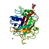
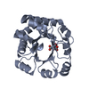
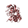
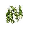
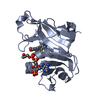
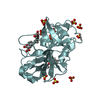
| Title | CATALYTIC DOMAIN OF PLASMINOGEN | ||||||
 Components Components | PLASMINOGEN | ||||||
 Keywords Keywords | HYDROLASE / MICROPLASMINOGEN / SERINE PROTEASE / ZYMOGEN / CHYMOTRYPSIN FAMILY | ||||||
| Function / homology |  Function and homology information Function and homology informationplasmin / trans-synaptic signaling by BDNF, modulating synaptic transmission / trophoblast giant cell differentiation / tissue remodeling / tissue regeneration / Signaling by PDGF / positive regulation of fibrinolysis / mononuclear cell migration / negative regulation of cell-cell adhesion mediated by cadherin / protein antigen binding ...plasmin / trans-synaptic signaling by BDNF, modulating synaptic transmission / trophoblast giant cell differentiation / tissue remodeling / tissue regeneration / Signaling by PDGF / positive regulation of fibrinolysis / mononuclear cell migration / negative regulation of cell-cell adhesion mediated by cadherin / protein antigen binding / Dissolution of Fibrin Clot / myoblast differentiation / labyrinthine layer blood vessel development / muscle cell cellular homeostasis / biological process involved in interaction with symbiont / Activation of Matrix Metalloproteinases / extracellular matrix disassembly / negative regulation of fibrinolysis / negative regulation of cell-substrate adhesion / apolipoprotein binding / positive regulation of blood vessel endothelial cell migration / fibrinolysis / Degradation of the extracellular matrix / platelet alpha granule lumen / serine-type peptidase activity / protein processing / Schaffer collateral - CA1 synapse / Regulation of Insulin-like Growth Factor (IGF) transport and uptake by Insulin-like Growth Factor Binding Proteins (IGFBPs) / kinase binding / blood coagulation / Platelet degranulation / protein-folding chaperone binding / extracellular matrix / protease binding / endopeptidase activity / blood microparticle / signaling receptor binding / serine-type endopeptidase activity / external side of plasma membrane / negative regulation of cell population proliferation / protein domain specific binding / glutamatergic synapse / enzyme binding / cell surface / proteolysis / : / extracellular exosome / extracellular region / plasma membrane Similarity search - Function | ||||||
| Biological species |  Homo sapiens (human) Homo sapiens (human) | ||||||
| Method |  X-RAY DIFFRACTION / SYNCHROTRON / Resolution: 2 Å X-RAY DIFFRACTION / SYNCHROTRON / Resolution: 2 Å | ||||||
 Authors Authors | Peisach, E. / Wang, J. / de los Santos, T. / Reich, E. / Ringe, D. | ||||||
 Citation Citation | Journal: Biochemistry / Year: 1999 Title: Crystal structure of the proenzyme domain of plasminogen. Authors: Peisach, E. / Wang, J. / de los Santos, T. / Reich, E. / Ringe, D. #1: Journal: Structure / Year: 1995Title: The Crystal Structure of the Catalytic Domain of Human Urokinase-Type Plasminogen Activator Authors: Spraggon, G.S. / Phillips, C. / Nowak, U.K. / Ponting, C.P. / Saunders, D. / Dobson, C.M. / Stuart, D.I. / Jones, E.Y. #2: Journal: Nat.Struct.Biol. / Year: 1998Title: The ternary microplasmin-staphylokinase-microplasmin complex is a proteinase- cofactor-substrate complex in action Authors: Parry, M.A. / Fernandez-Catalan, C. / Bergner, A. / Huber, R. / Hopfner, K.P. / Schlott, B. / Guhrs, K.H. / Bode, W. #3: Journal: Science / Year: 1998Title: Crystal structure of the catalytic domain of human plasmin complexed with streptokinase Authors: Wang, X. / Lin, X. / Loy, J.A. / Tang, J. / Zhang, X.C. | ||||||
| History |
|
- Structure visualization
| Structure viewer | Molecule: MolmilJmol/JSmol |
|---|
- Downloads & links
-Download
| PDBx/mmCIF format | 1qrz.cif.gz | 205 KB | Display | PDBx/mmCIF format |
|---|---|---|---|---|
| PDB format | pdb1qrz.ent.gz | 164.6 KB | Display | PDB format |
| PDBx/mmJSON format | 1qrz.json.gz | Tree view | PDBx/mmJSON format | |
| Others |  Other downloads Other downloads |
-Validation report
| Arichive directory | https://data.pdbj.org/pub/pdb/validation_reports/qr/1qrzftp://data.pdbj.org/pub/pdb/validation_reports/qr/1qrz | HTTPS FTP |
|---|
-Related structure data
| Similar structure data |
|---|


-Links
 PDBj
PDBj


- Assembly
| Deposited unit | 
| ||||||||
|---|---|---|---|---|---|---|---|---|---|
| 1 | 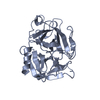
| ||||||||
| 2 | 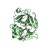
| ||||||||
| 3 | 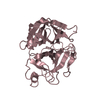
| ||||||||
| 4 | 
| ||||||||
| Unit cell |
|
-Components
| #1: Protein | Mass: 27020.016 Da / Num. of mol.: 4 / Fragment: CATALYTIC DOMAIN / Mutation: M585Q, V673M, M788L Source method: isolated from a genetically manipulated source Source: (gene. exp.) Homo sapiens (human) / References: UniProt: P00747#2: Water | ChemComp-HOH / |  Mass: 18.015 Da / Num. of mol.: 509 / Source method: isolated from a natural source / Formula: H2O Mass: 18.015 Da / Num. of mol.: 509 / Source method: isolated from a natural source / Formula: H2OHas protein modification | Y | |
|---|
-Experimental details
-Experiment
| Experiment | Method: X-RAY DIFFRACTION / Number of used crystals: 1 |
|---|
- Sample preparation
| Crystal | Density Matthews: 2.14 Å3/Da / Density % sol: 42.53 % | |||||||||||||||||||||||||||||||||||
|---|---|---|---|---|---|---|---|---|---|---|---|---|---|---|---|---|---|---|---|---|---|---|---|---|---|---|---|---|---|---|---|---|---|---|---|---|
| Crystal grow | Temperature: 277 K / Method: vapor diffusion, hanging drop / pH: 5.6 Details: PEG 4000, ISOPROPANOL, CITRATE, pH 5.6, VAPOR DIFFUSION, HANGING DROP, temperature 277K | |||||||||||||||||||||||||||||||||||
| Crystal grow | *PLUS pH: 6.2 | |||||||||||||||||||||||||||||||||||
| Components of the solutions | *PLUS
|
-Data collection
| Diffraction | Mean temperature: 100 K |
|---|---|
| Diffraction source | Source: SYNCHROTRON / Site: NSLS  / Beamline: X12B / Wavelength: 0.9924 / Beamline: X12B / Wavelength: 0.9924 |
| Detector | Type: ADSC QUANTUM 4 / Detector: CCD / Date: Oct 14, 1998 |
| Radiation | Protocol: SINGLE WAVELENGTH / Monochromatic (M) / Laue (L): M / Scattering type: x-ray |
| Radiation wavelength | Wavelength: 0.9924 Å / Relative weight: 1 |
| Reflection | Resolution: 2→100 Å / Num. all: 63217 / Num. obs: 63217 / % possible obs: 99.3 % / Observed criterion σ(F): 0 / Observed criterion σ(I): 0 / Redundancy: 6.9 % / Biso Wilson estimate: 3.6 Å2 / Rmerge(I) obs: 0.165 / Net I/σ(I): 11 |
| Reflection shell | Resolution: 2→2.07 Å / Redundancy: 2.7 % / Rmerge(I) obs: 0.631 / % possible all: 98 |
| Reflection | *PLUS Highest resolution: 2 Å / Num. measured all: 199470 |
- Processing
| Software |
| ||||||||||||||||||||||||||||||||||||||||||||||||||||||||||||||||||||||||||||||||
|---|---|---|---|---|---|---|---|---|---|---|---|---|---|---|---|---|---|---|---|---|---|---|---|---|---|---|---|---|---|---|---|---|---|---|---|---|---|---|---|---|---|---|---|---|---|---|---|---|---|---|---|---|---|---|---|---|---|---|---|---|---|---|---|---|---|---|---|---|---|---|---|---|---|---|---|---|---|---|---|---|---|
| Refinement | Resolution: 2→35 Å / Rfactor Rfree error: 0.005 / Data cutoff high absF: 95413.39 / Data cutoff low absF: 0 / Isotropic thermal model: RESTRAINED / Cross valid method: THROUGHOUT / σ(F): 0 / σ(I): 0 / Stereochemistry target values: ENGH & HUBER / Details: MAXIMUM LIKELIHOOD TARGET FUNCTION
| ||||||||||||||||||||||||||||||||||||||||||||||||||||||||||||||||||||||||||||||||
| Solvent computation | Solvent model: FLAT MODEL / Bsol: 24.27 Å2 / ksol: 0.311 e/Å3 | ||||||||||||||||||||||||||||||||||||||||||||||||||||||||||||||||||||||||||||||||
| Displacement parameters | Biso mean: 19.1 Å2
| ||||||||||||||||||||||||||||||||||||||||||||||||||||||||||||||||||||||||||||||||
| Refine analyze |
| ||||||||||||||||||||||||||||||||||||||||||||||||||||||||||||||||||||||||||||||||
| Refinement step | Cycle: LAST / Resolution: 2→35 Å
| ||||||||||||||||||||||||||||||||||||||||||||||||||||||||||||||||||||||||||||||||
| Refine LS restraints |
| ||||||||||||||||||||||||||||||||||||||||||||||||||||||||||||||||||||||||||||||||
| LS refinement shell | Resolution: 2→2.13 Å / Rfactor Rfree error: 0.018 / Total num. of bins used: 6
| ||||||||||||||||||||||||||||||||||||||||||||||||||||||||||||||||||||||||||||||||
| Xplor file |
| ||||||||||||||||||||||||||||||||||||||||||||||||||||||||||||||||||||||||||||||||
| Software | *PLUS Name: CNS / Version: 0.5 / Classification: refinement | ||||||||||||||||||||||||||||||||||||||||||||||||||||||||||||||||||||||||||||||||
| Refinement | *PLUS σ(F): 0 / % reflection Rfree: 4.9 % | ||||||||||||||||||||||||||||||||||||||||||||||||||||||||||||||||||||||||||||||||
| Solvent computation | *PLUS | ||||||||||||||||||||||||||||||||||||||||||||||||||||||||||||||||||||||||||||||||
| Displacement parameters | *PLUS Biso mean: 19.1 Å2 | ||||||||||||||||||||||||||||||||||||||||||||||||||||||||||||||||||||||||||||||||
| Refine LS restraints | *PLUS
| ||||||||||||||||||||||||||||||||||||||||||||||||||||||||||||||||||||||||||||||||
| LS refinement shell | *PLUS Rfactor Rfree: 0.371 / % reflection Rfree: 5 % / Rfactor Rwork: 0.301 |
